VOTW: Tendon to Business

 ·   · 

HPI

A 25M with an unremarkable PMH presents with 3 days of atraumatic left medial ankle pain, redness, and swelling with no other associated symptoms. He is unable to bear weight or ambulate today due to pain.

Physical exam reveals an erythema and tenderness just posterior to the left medial malleolus with no palpable ankle joint effusion.

Ultrasound

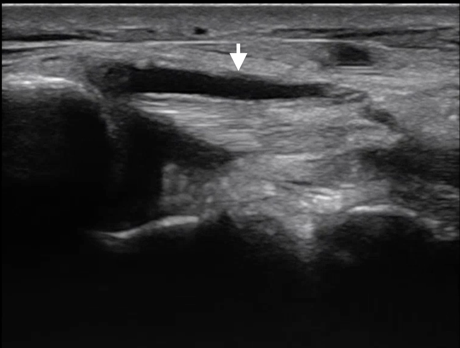
POCUS of the left lower extremity demonstrated no DVT in the femoral or popliteal veins, no abscess in the overlying soft tissue, and no joint effusion in the ankle. Fluid was noted surrounding the posterior tibial tendon, shown below in two planes.

The unaffected contralateral tendon is shown for comparison.

Characteristic findings of tendinitis on ultrasound include hypoechoic areas of inflammation, tendon thickening, or peritendinous fluid. POCUS is also useful to assess for other conditions like tendon rupture or tears.

Case Conclusion

CT imaging showed thickening of the posterior tibial tendon with surrounding edema suggestive of tendinopathy with no evidence of abscess. Antibiotics were started for possible infectious etiology. Patient was ambulatory after pain control and was given a referral for orthopedic followup.

Happy scanning!


VOTW: Do you know the muffin man?

 ·   · 

Case: 57 yoF with PMHx of osteoarthritis presented to the ED with right knee pain. A bedside US was performed that showed bulging of the right medial meniscus with surrounding edema Image 1, videos 1&2), consistent with a tear in the medial meniscus.

Meniscal Tear

How to assess the meniscus:

  1. Have the patient flex their knee slightly to 20-30 degrees (you can prop their knee up with a rolled towel) 

  2. Use the linear probe and place it longitudinally along the medial aspect of the knee 

  3. Identify the medial collateral ligament (MCL), which will appear as a hyperechoic and fibrillar structure, extending from the medial femoral condyle to the proximal tibia

  4. The meniscus will appear as a triangular structure that sits between the femur and tibia, under the MCL

5. A meniscal tear is identified with a well-defined anechoic or hypoechoic area surrounding the meniscus. It can cause extrusion of the meniscus as a result of surrounding edema, described as a "muffin top". There may also be increased vascularity in the surrounding area when color flow doppler is applied

Note: The same process can be repeated on the lateral aspect of the knee to assess the lateral meniscus (image 3).

Case conclusion: While the patient had pain with flexion, she was able to ambulate independently. She was discharged with orthopedic follow-up and conservative measures.

Happy scanning! 

Ariella Cohen


References: 

https://theultrasoundsite.co.uk/ultrasound-case-studies/

https://ultrasoundpaedia.com/knee-normal/

https://www.nysora.com/ultrasound-of-the-musculoskeletal-system/chapter14-knee-preview/


VOTW: Big Flex

77 yoF presented to the ED with left middle finger pain, swelling, and discoloration of ~1 week proximally (image 1). 

Image 1: digit swelling and abscess

She was tachycardic and febrile on arrival. Physical exam revealed ¾ Kanavel signs (flexion at rest, pain with passive extension, fusiform swelling of the digit). Ultrasound was performed using a water bath (image 2).

Image 2: water bath technique

POCUS findings concerning for flexor tenosynovitis include a hypoechoic peritendunous effusion and a thickened synovial sheath that may be hyperemic.

Video 1/Image 3: shows fluid surrounding the flexor tendon in short axis.

Image 3: Short axis of flexor tendon with surrounding fluid

Video 2/Image 4: shows a long axis view of the finger with fluid in the pre-tendon area and surrounding edema.

Image 4: Long axis of flexor tendon with abscess/edema

Case conclusion: orthopedics was called and performed a bedside I&D. They were able to express “copious amounts of purulent fluid” but did not appreciate pus along the tendon sheath itself. The patient was admitted for IV abx and is getting daily wound checks by orthopedics.

Note: While the Kanavel signs and ultrasound are useful diagnostic aids, they are non-specific and should not be used as a rule-out test. Remember that many inflammatory processes will often create edema that appears hypoechoic on ultrasound.

Happy scanning!

- Ariella Cohen

References:

https://www.emdocs.net/ultrasound-probe-pocus-for-flexor-tenosynovitis/

https://www.researchgate.net/figure/Ultrasound-appearance-of-normal-flexor-tendon-sheath-and-tenosynovitis-a-Normal_fig1_51104450