Analgesia for Acute Anterior Shoulder Dislocation Reduction

 ·   · 

Hello all,

For this Trauma Tuesday imagine you're a 1945 WWII nurse transported back in time to 1743 Scotland and a Jacobite warrior dislocates his shoulder. You need to reduce it but all they have for pain control is Scottish whiskey and if you're lucky, something called Laudanum. (So goes the plot of Outlander)

Laudanum is a mixture of opium and ethanol which is old school procedural sedation and analgesia (PSA) that is equivalent to fentanyl and versed that we use in the modern day. It is definitely a tried-and-true combo that has crossed into the 21st century but how does it fair against intra-articular lidocaine (IAL)?

First off, anterior shoulder dislocations is the most common shoulder dislocation ~95%, posterior shoulder dislocation ~5%, and inferior shoulder dislocation <1%.

In these images, the red arrow is pointing at the glenoid fossa and the blue arrow is pointing at the humeral head which should be in contact with the glenoid fossa.

To inject Intra-articular lidocaine first locate the landmarks, since this is an anterior shoulder dislocation the posterior approach to the glenohumeral joint and subacromial space may be the easiest as that space is widened due to the dislocation. 

Have the patient sit with their arm resting at their side if possible. Palpate the posterior indent between the head of the humerus and acromion, using a 18G needle with 20 mL of 1% lidocaine, insert the needle 2-3cm inferior and medial to the posterolateral corner of the acromion and direct it anteriorly towards the coracoid process when aiming for the glenohumeral joint. When aiming for the subacromial space, direct it laterally while aiming for the anterolateral acromion corner. However, any space between these two areas will be sufficient for an intra-articular block with a dislocation. An 18G needle should sink into the space and the plunger should push with no resistance if you are in the correct space. 

One study did a systematic review and meta-analysis of 12 RCTs comparing IAL to PSA (with any agent) for closed reduction of acute anterior shoulder dislocation for patients > 15 years of age.

Results:

  • No difference in reduction success between IAL and PSA. (83.8% vs. 91.4%)

  • Fewer adverse events occurred in the IAL group compared to the PSA group. (1.3% vs. 20.8%) 

  • Mean ED length of stay was significantly shorter in the IAL group compared to the PSA group. (mean difference = − 1.48 h)

  • No difference in pain score after anesthesia and before reduction in the IAL group compared to the PSA group. (mean difference = − 0.04)

  • Procedural time was significantly longer in the IAL group compared to PSA (mean difference = 8 min)

  • No statistically significant difference in ease of reduction in the IAL group compared to PSA. (54.5% vs. 71.8%)

  • Patient satisfaction was significantly decreased in the IAL group compared to PSA. (70.5% vs. 90.4%)

TL;DR

IAL seems to have similar effectiveness as PSA in the successful reduction of anterior shoulder dislocations in the ED with fewer adverse events, shorter ED length of stay, and no difference in pain scores, or ease of reduction. However, IAL had longer procedural times and decreased patient satisfaction which is something to consider. Nonetheless, IAL may be an effective alternative to procedural sedation for reducing anterior shoulder dislocations, particularly when IV sedation is contraindicated or not feasible.

https://rebelem.com/intra-articular-lidocaine-vs-procedural-sedation-and-analgesia-for-closed-reduction-of-acute-anterior-shoulder-dislocation/

https://wikem.org/wiki/Shoulder_dislocation

https://www.shoulderdoc.co.uk/article/1485

Sithamparapillai, A, Grewal, K,Thompson, C, Walsh, C, McLeod, S. Intra-Articular lidocaine versus intravenous sedation for closed reduction of acute anterior shoulder dislocation in the Emergency Department: a systematic review and meta-analysis. Canadian Journal of Emergency medicine. October 2022. PMID: 36181665

 · 
Share

Pong Pong Tree

 ·   · 

Hello everyone!

For those of you who don't know me, I am Karen, a the third year resident who will be staying on as one of the ultrasound fellows next year (along with the amazing Dan Ye, who can forget his great recaps of The Pitt), I'm also one of the four chiefs following our previous admin resident and chief Kaitlyn DeStefano (whose POTDs made us laugh and cry). I have big shoes to fill with these two tough acts to follow, but I will work hard to bring fun and educational POTDs!

Continuing on The Pitt bandwagon, I love watching shows, from romances and drama, to murder mysteries and thrillers, to Kdramas and anime; I am a huge consumer of media for better or for worse. Some of my favorites are Outlander, Downton Abbey, Dark, Black Mirror, Squid Game, Attack on Titan, When Life Gives You Tangerines, of course The Pitt and so many more...

Anyways, since yesterday was the finale of HBO's "White Lotus" I wanted to talk about the Pong pong tree. (Spoilers!)

The Pong Pong tree (Cerbera odollam)—also known as the “suicide tree” is native to India and Southeast Asia. This plant contains cerberin, a cardiac glycoside similar to digoxin that disrupts cardiac activity by inhibiting the Na⁺/K⁺- ATPase pump, which can lead to fatal arrhythmias. Cerberin poisoning is difficult to detect and has been used in homicides due to its tasteless nature and delayed onset. There is no specific antidote, though digoxin-specific Fab fragments (DIGIFab) has been used.

There has been only 2 known cases of C. odollam poisoning and 1 fatality reported in the USA:

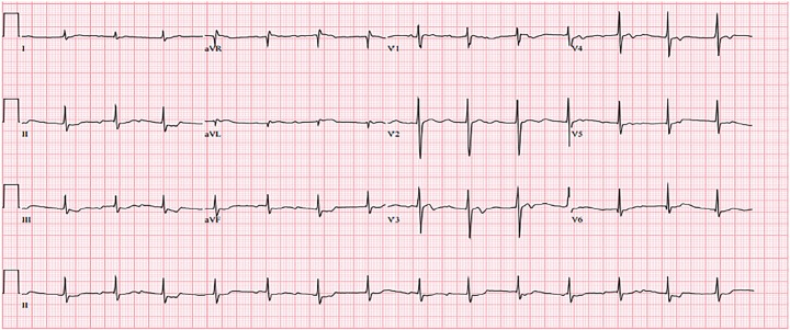
A 22-year-old pre-operative transgender man-to-woman patient ingested seeds from the Cerbera odollam tree as a suicide attempt. She presented to the emergency department with nausea, vomiting, chest pain, and dizziness approximately 7 hours after ingesting the seeds that she bought online after reading about suicide. 

Initial electrocardiogram (ECG) showed second-degree heart block with 2:1 atrioventricular (AV) conduction and ST-segment depression with biphasic T-waves and initial serum potassium was 5.2mEq/L:

So the team administered atropine, digoxin-Fab fragments, and supportive care with improvement to sinus rhythm with first-degree AV block with persistent ST-segment depression and biphasic T-waves:

However, 2 hours later her condition deteriorated, progressing to high-degree AV block:

And subsequent cardiac arrest 30 minutes later with repeat K of 5.7mEq/L.

Despite additional doses of digoxin-specific antibody fragments (20 vials in total), lipid emulsion 20% (100 ml), right femoral CVC, and 2 hours of aggressive ACLS resuscitative efforts the patient was pronounced dead approximately 12 hours post-ingestion.

This case delineates the severe toxicity associated with C. odollam seed ingestion and highlights the challenges in managing such poisonings, even with advanced supportive measures. We should all be aware of the potential morbidity and mortality linked to this plant toxin and be prepared for aggressive resuscitative interventions.

Although the USA has only had 2 known cases of C. odollam poisoning, will a television show introducing this plant open the gates for more? Hopefully not, but as always, we will be prepared.

Misek R, Allen G, LeComte V, Mazur N. Fatality Following Intentional Ingestion of Cerbera odollam Seeds. Clin Pract Cases Emerg Med. 2018 Jun 12;2(3):223-226. doi: 10.5811/cpcem.2018.5.38345. PMID: 30083638; PMCID: PMC6075506. https://pmc.ncbi.nlm.nih.gov/articles/PMC6075506/

Karen Wong, MD

Emergency Medicine Chief Resident

KaWong@maimo.org

 · 
Share

Pneumomediastinal Tamponade

Today's POTD came as a request from the Maimonides Attending Group Chat! This was a request after the Pitt, so taking a play out of Dr. Dan Ye's book to talk about case that is described in the latest episode. 


So without further adue lets look at some pneumomediastium specifically pneumomediastium causing tamponade.


Pneumomediastium: air present in the mediastinum, usually this occurs from air extravastaing from within the airways/lungs or esophagus and migrates into the mediastium. This air then dissects the cervical subcutanous tissues, epidural space, pericardium, and/or peritoneal cavity. 


Pneumomediastium is either typically spontaneous, rare, and typically self limited. Consider risk factors including smoking/tobacco use, recreational drug inhalation. Other causes include intrinsic lung and airway causes including asthma, COPD, bronchiectasis, COPD, ILDz, lung cancer, foreign body, increased intrathoracic pressure (forceful sneezing), increased intravagal tone (such as with childbirth, excessive vomiting, or strenous physical activities). Other iatrogenic cauases include: endoscopy, intubation, central line placement, thoracostomy, or chest/abdominal surgeries, and probably most commonly: traumatic causes: blunt trauma, penetrating trauma, or blast injuries. 


In the Pitt, **SPOILER ALERT**, the patient was shot in the chest and had a penetrating trauma.


Spontaneous pneumomediastum can also present with younger males of tall structure with low body mass. This spontaneous pneumomediastium occurs because of Macklin phenomenon: increased intra-aveolar pressure --> alveolar rupture --> air dissects into peribronchial and perivascular sheaths --> air progresses itno mediastium and surrounding tissues. 


Presentation: 

Most of the time this occurs with retrosternal chest apin that may radiate into the back or neck. 

On physical exam, these patients present with subcutaneous emphysema 

Other signs and symptoms include: rhinolalia (nasal tone of speech), dysphonia, neck swelling, hoarsness, tachycardia or tachypnea 


Diagnosis: 

Diagnosis is made usually on xray, and clinical diagnosis. 

On CXR you may see subcutaneous emphysema, elevated thymus (in peds patients), air around the pulmonary arteries, V shape between the descending aorta and left hemidiaphragm, double bronchial wall, or pleural effusion.


In pneumomediastium causing tamponade, these patients may have explained hypotension. Normally when we think about tamponade, we are able to be clused in with ultrasound findings, pneumomediastium on ultrasound may be seen as the "air gap" sign which would appear as sonographic echos, usually seen with M mode, that obsecure the cardiac structures underneath. If you have ever tried to place an ultrasound probe on a patient's chest with subq emphysema, you see nearly nothing, so this is definitiely more a clinical diagnosis to consider with unexplained hypotension in someone with subq emphysema. 


Tension pneumomediastinum/tamponade leads to compression of great vessels and compromises venous return --> hypotension and hypoxemia 


Management of tension pneumomediastinum that causes tamponade physiology: 

In the show, the patient got bilateral "blowholes": performed by a 2 cm incision infraclavicular through the skin and prepectoral fascia 

Other methods described include b/l chest tubes, placement of penrose drains into the neck 


Otherwise pneumomediastium is treated with supportive care and usually resolves on its own. 


Until next time friends!

Fleming AM, Zambetti BR, Valaulikar GS. Bedside Mediastinotomy for Tension Pneumomediastinum With Tamponade in COVID-19. Ann Thorac Surg. 2021 Oct;112(4):e265-e266. doi: 10.1016/j.athoracsur.2021.01.032. Epub 2021 Jan 30. PMID: 33529601; PMCID: PMC8402946


Iteen AJ, Bianchi W, Sharman T. Pneumomediastinum. [Updated 2023 May 1]. In: StatPearls [Internet]. Treasure Island (FL): StatPearls Publishing; 2025 Jan-. Available from: https://www.ncbi.nlm.nih.gov/books/NBK557440/

Sciortino CM, Mundinger GS, Kuwayama DP, Yang SC, Sussman MS. Case report: treatment of severe subcutaneous emphysema with a negative pressure wound therapy dressing. Eplasty. 2009;9:e1. Epub 2009 Jan 7. PMID: 19198645; PMCID: PMC2627309.

https://radiopaedia.org/articles/pneumomediastinum?lang=us

 · 
Share