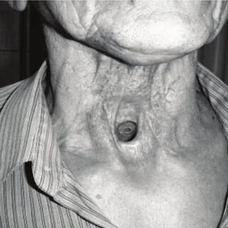
POTD: Tracheostomy and Laryngectomy Emergencies

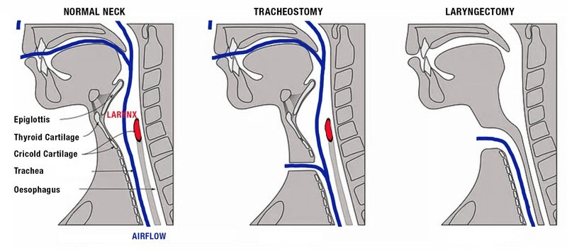
What is the difference between tracheostomy and laryngectomy?

  • Laryngectomies do not have a connection between the oropharynx and upper airway and cannot be intubated via mouth or nose.

  • Tracheostomy complications are more common and will be the focus of today's emails. Laryngectomies have become a rarer procedure. 

Tracheo-innominate artery fistula

    • Presentation

      1. Massive hemorrhage from tracheostomy, usually within 3-4 weeks of placement

    • Workup

      • CTA, bronchoscopy, local exploration

      • Note, there is high risk of recurrence so workup should be done even if bleeding has stopped.

    • Management

      • Emergent consult to ENT, general surgery, or vascular surgery.

      • Obtain access, transfuse blood products, reverse anticoagulants.

      • External compression over sternal notch

      • Compress innominate artery with overinflated cuff.

        • If tube is cuffed, overinflate cuff with up to 50ml air to compress innominate artery.

        • If tube is uncuffed,

          • If tracheostomy > 7 days, exchange with cuffed tube and overinflate

          • If tracheostomy < 7 days, exchange under endoscopy or over a bougie if endoscope is not available.

      • Digital compression of innominate artery (Utley Maneuver)

        • Insert ET tube via mouth or stoma deep to innominate artery, then insert finger into stoma and digitally compress innominate artery.

    • Disposition

      • Low threshold for admission to step-down or ICU setting.

Obstruction

    • Presentation

      • Respiratory distress with minimal airflow around tracheostomy, sometimes with clear mucus plugging.

      • High risk in small tube size.

    • Workup

      • Attempt to pass flexible suction catheter.

      • Can visualize via endoscopy

    • Management (stop after each step to assess resolution of obstruction)

      • Oxygen via face mask and via tracheostomy.

      • Remove external devices (i.e speaking valves, dressings, obturators)

      • Remove inner cannula

      • Suction outer cannula with flexible suction

      • Deflate cuff to allow air flow around tube, assuming tube is obstructed

      • Remove tracheostomy

      • BVM via face or stoma. Be sure to occlude the end that you are not ventilating though to prevent air leak.

      • Reintubate if needed. Orotracheal intubation preferred, but can also intubate via stoma if needed (i.e. upper airway blocked by tumor)

        1. 6-0 tube or smaller if intubating via stoma.

    • Disposition

      • Discharge with ENT follow-up if obstruction if cleared, stable respiration

      • Admit to ICU if significant hypoxic event or tenuous airway patency 2/2 recurrent obstruction.

      • Admit to stepdown/floors otherwise

Decannulation

    • Presentation

      1. Partial or complete displacement of tracheostomy tube, w/ poor air movement.

    • Workup

      • Attempt to pass flexible suction catheter.

      • Visualize with endoscopy.

    • Management

      • High flow oxygen to face and stoma

      • If tracheostomy < 7 days old, replace only under direct visualization with endoscope. If no endoscopy, then orally intubate.

      • If tracheostomy > 7 days old, place new tracheostomy tube in stoma. If there is resistance, reattempt with a downsized tube.

      • Assess for subcutaneous emphysema, which can indicate you are in a false-passage or tracheal injury.

      • Monitor with capnography

    • Disposition

      • Admit if difficult reintubation, requiring downsizing of tube or increased airway secretions

      • Admit to ICU if pt required orotracheal intubation, significantly increased suction burden, or AMS requiring ICU monitoring.

      • Discharge with ENT followup if patient had mature tracheostomy with tube replaced easily and placement is confirmed by capnography/bedside endoscopy.

Laryngectomy patients

  • Less common procedure now

  • No anatomic connection from trachea to face.

    • Cannot be nasally or orally intubated.

    • Nasal cannula or face mask will not deliver gas to lungs.

  • Some patients may have tracheoesophageal puncture voice prosthesis that can dislodge and become aspirated. You should be able to see this in the stoma.

  • Workup

    • CXR, +/- CT chest for foreign bodies, alternate lung pathologies

    • Consider CT neck w/ contrast, bronchoscopy, cultures for infectious workup

  • Management

    • O2 via stoma only. Do not intubate via mouth or nose.

    • ENT consult for infections, prosthesis aspirations

  • Disposition

    • Discharge home if reversible etiology (i.e. obstruction/secretions) that has been resolved without complication. May discharge minor soft tissue infections in consultation w/ ENT

    • Admit aspirated TEP prosthesis for retrieval and monitoring, or if cause of dyspnea is unclear.

References

Manning Sara, Bontempo Laura. Complications of Tracheostomies and Laryngectomies. In: Mattu A and Swadron S, ed. CorePendium. Burbank, CA: CorePendium, LLC. https://www.emrap.org/corependium/chapter/reckOdDn9Ljn7sBLy/Complications-of-Tracheostomies#h.5xyzow82ssmf. Updated September 21, 2023. Accessed December 16, 2023.

 · 
Share

POTD: Influenza

Clinical presentation

  • Sore throat, fevers, chills, myalgias

  • If severe, will result in hypoxemia and tachypnea.

When to hospitalize

  • Significant dehydration

  • If the patient appears septic: respiratory distress, hypoxemia, impaired cardiopulmonary function, AMS.

Imaging

  • US/CXR show patchy bilateral infiltrates.

  • CT may show bronchial wall thickening, tree-in-bud nodules, multifocal consolidations.

Testing

  • Flu PCR has ~ 90% sensitivity, though is dependent on quality of sample (did the swab go deep enough)

  • Tracheal aspirate or BAL if intubated (gold standard)

  • Procalcitonin. Influenza generally doesn’t increase procalcitonin à be more suspicious of a bacterial cause.

  • General sepsis/pneumonia labs: blood and sputum cultures, MRSA PCR, urine

When to suspect a bacterial superinfection

  • Imaging with lobar consolidations, cavitations, or significant pleural effusion suggests superimposed bacterial PNA, or other diagnosis.

  • A biphasic illness: when the patient initially improves, then deteriorates again.

  • Copious sputum production (generally not a feature of influenza)

Is there a role for antivirals?

  • The evidence is iffy. Cochrane review 2014 of 44 trials, 24,000 patients showed only modest benefits1

    • Reduction of symptoms by ~ 0.5 days average.

    • Does not reduce hospitalization or development of pneumonia.

  • 1st line: Tamiflu (oseltamivir) 75mg BID x5 days (10 days if critically ill). PO only.

    • Most effective within the first 48 hours if the patient is critically ill, give it regardless of illness duration.

  • 2nd line: Peramivir in pt who cannot tolerate oral therapy.

Is there a role for antibiotics?

No… but also yes. There is bacterial superinfection in 1/3rd of patients, and it is generally difficult to exclude bacterial pneumonia until cultures return.

  • Antibiotic choices include beta-lactam + macrolide + MRSA coverage.

  • Beta-lactam:

    • Generally, ceftriaxone.

    • Broaden to pseudomonal coverage for your HCAP/VAP/immune compromised patients, same as your other pneumonias.

  • Macrolide

    • Instead of azithromycin, consider clarithromycin, which has direct antiviral activity against influenza2.

  • MRSA coverage

    • High prevalence of MRSA superinfection among influenza pneumonia 3

    • Linezolid is superior to vancomycin for MRSA pneumonia, unless there is a contraindication4.

A key difference in resuscitation versus traditional sepsis

  • Most mortality in influenza pneumonia is secondary to ARDS5. Avoid large volume resuscitation. I.e. do not follow the Surviving Sepsis 30cc/kg recommendation.

  • For mild hypotension, consider low dose pressors instead of large volume fluid resuscitation.

  • If the patient is normotensive, great. You don’t need to resuscitate blood pressure.


References

1.           Jefferson T, Jones MA, Doshi P, et al. Neuraminidase inhibitors for preventing and treating influenza in adults and children. Cochrane Database Syst Rev. 2014;(4). doi:10.1002/14651858.CD008965.pub4

2.           Yamaya M, Hatachi Y, Kubo H, Nishimura H. Clarithromycin inhibits pandemic A/H1N1/2009 influenza virus infection in human airway epithelial cells. Eur Respir J. 2012;40(Suppl 56). Accessed December 11, 2023. https://erj.ersjournals.com/content/40/Suppl_56/P4364

3.           Uyeki TM, Bernstein HH, Bradley JS, et al. Clinical Practice Guidelines by the Infectious Diseases Society of America: 2018 Update on Diagnosis, Treatment, Chemoprophylaxis, and Institutional Outbreak Management of Seasonal Influenzaa. Clin Infect Dis Off Publ Infect Dis Soc Am. 2019;68(6):e1-e47. doi:10.1093/cid/ciy866

4.           Wunderink RG, Niederman MS, Kollef MH, et al. Linezolid in methicillin-resistant Staphylococcus aureus nosocomial pneumonia: a randomized, controlled study. Clin Infect Dis Off Publ Infect Dis Soc Am. 2012;54(5):621-629. doi:10.1093/cid/cir895

5.           Severe influenza. EMCrit Project. Accessed December 11, 2023. https://emcrit.org/ibcc/influenza/

 · 
Share