ED Thoracotomy

It’s a new academic year and we are going to start off with some trauma! Today, we are going to talk about ED thoracotomy. It’s basically surgery but in the ED.

First of all, What is ED thoracotomy? It is a Hail Mary attempt in patients with penetrating chest injuries with signs of life in the field and suffer a subsequent witnessed cardiac arrest to open the chest and temporize a life threatening wound. It can be used to relieve cardiac tamponade, control hilar bleeding, cross clamp the descending aorta, or repair a myocardial laceration, all while on the way to the OR for definitive repair.

Success rate and survival is very poor for patients when thoracotomy is performed for blunt trauma, about 2% survival for patients in shock and less than 1% for patients with no vital signs. Conversely, in penetrating wounds, while still poor, 15% of all patients survived.

Guidelines have been established, the so called East and West guidelines, to help balance the already poor odds of survival with the risks of exposing health care providers to blood borne pathogens and salvaging patients with high odds of anoxic brain injury.

Western Trauma Association guidelines: consider ED thoracotomy in patients arriving with blunt trauma and < 10 minutes of CPR or penetrating trauma and < 15 minutes of CPR.

Eastern Association for the Surgery of Trauma: strongest recommendation to perform ED thoracotomy in patients with initial signs of life after penetrating thoracic injury who now present pulseless.

thoracotomy.png

Other commonly quoted numbers. Also consider thoracotomy when you have unresponsive hypotension SBP < 70 despite aggressive resuscitation, chest tube output > 1500cc’s of blood within 24 hours, or when there is > 200/250 cc/hr output of blood after tube thoracostomy for 2-4 consecutive hours.

What you need: A well-staffed and properly trained team that includes also includes a trauma surgeon. Lots of PPE. Thoracotomy tray (rib spreader, #10 or #21 scalpel, scissors, foreceps, vascular clamps, curved artery forceps, needle driver, internal defibrillation paddles, skin stapler, sutures). I would also add a foley catheter as it can be inserted into the heart and the balloon inflated to stabilize a laceration while sutures are being placed.

How is it done? These are the basic steps. Secure the airway and insert an NGT, it can help distinguish the aorta which lies posterior to esophagus. Then, start with a L sided approach, place a chest tube on the R side concurrently. Incise from the sternum to the posterior axillary line at the 4/5th intercostal space, cutting through skin/soft tissue and muscle in one go. Spread the ribs with the rachet bar down. Pick up the pericardium and open it. Inspect myocardium for lacerations. Cardiac massage, internal defibrillation, and intracardiac epinephrine can be done. The aorta can be cross clamped for up to 30 minutes if hypotension still persists. If no evidence of L sided injury, extend to the R side (clam shell).


Hope this helps next time you get a note about a stab or GSW, active CPR, 3 minutes out!

Sources

https://emcrit.org/emcrit/procedure-of-thoracotomy/

https://i0.wp.com/emcrit.org/wp-content/uploads/2012/10/CJLDP_bW8AAVQ2o.png-large.png

https://westerntrauma.org/documents/PublishedAlgorithms/WTACriticalDecisionsResuscitativeThoracotomy.pdf

https://www.east.org/education/practice-management-guidelines/emergency-department-thoracotomy

https://jamanetwork.com/journals/jamasurgery/fullarticle/391389

https://wikem.org/wiki/Thoracotomy

West guidelines algorithm.jpg
 · 
Share

Diplopia

 ·   · 

Where to start?

First, figure out is it monocular or binocular diplopia?

Monocular: diplopia persists with one eye closed

Almost exclusively an eye problem.  It is almost always benign and most often due to refractive error. Give ophthalmology referral and no further imaging is indicated unless warranted by other signs or symptoms.

Binocular: diplopia resolves with one eye closed

Often due to neuromuscular dysfunction causing misalignment of gaze. 

This requires further evaluation in the ED to rule out acute neurological injury.


Next, is the diplopia isolated or associated with other neurologic signs or symptoms?

Do a thorough neurological exam. 

If you find any other neurological symptoms or signs, consider a stroke code/consult and get CT/MRI.  

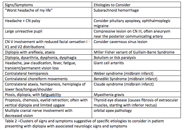
Think of the following table of diagnoses for patients presenting with diplopia and focal neurological deficits, and order the appropriate tests and imaging needed:

Table for Diplopia DDx.png

If there is an isolated palsy, try to localize the lesion.

Ocular Palsies.png

Isolated 3rd nerve palsy: may be due to a cerebral aneurysm, especially if affecting the pupil, and must be emergently evaluated with noncontrast head CT and CTA

Isolated 4th nerve palsy: usually present with vertical or diagonal diplopia, usually due to trauma or idiopathic; be sure to check for cerebellar signs because the 4th nerve exits on the dorsum of the brainstem and can be compressed by a posterior fossa tumor. If no cerebellar signs or trauma, can be discharged with an ophthalmology referral.

Isolated 6th nerve palsy: present with horizontal diplopia, usually idiopathic, check for bilateral 6th nerve palsies as this can be a sign of a tumor (some sources state an isolated 6th nerve palsy should also be evaluated with head imaging prior to dispo). 

For horizontal diplopia, evaluate for internuclear ophthalmoplegia, and if present, get an MRI. Various causes, including multiple sclerosis. 

In children, there is a higher prevalence of malignancy (vs microvascular insult) so they always require head imaging.

Now what?

The need for neuroimaging in diplopia will depend on the palsy the patient has as well as the presence of other abnormalities in the history or physical exam. 

Isolated 4th nerve palsies and 6th nerve palsies can be referred to a specialist for evaluation and do not require imaging in the ED (except as outlined above).

The presence of a 3rd nerve palsy, multiple concomitant palsies, evidence of papilledema, infection, trauma, or cavernous sinus thrombosis require urgent imaging in the ED.

  • 3rd cranial nerve involvement: CTA

  • Increased ICP: CT head

  • Associated neuro deficits/complex motility disorders: CT head, preferably MRI

  • Suspected infection: CT head/orbits with contrast

  • Suspected Cavernous Sinus Thrombosis: CTV, followed by MRV if CTV negative

Patients over age 50 with diplopia should have inflammatory markers ordered.
Diplopia merits urgent referral to an ophthalmologist or neurologist once immediate, intervenable causes have been ruled out.

Sources

http://www.emdocs.net/diplopia-evaluation-and-management/

https://www.nuemblog.com/blog/double-vision

https://crashingpatient.com/wp-content/uploads/2018/06/diplopia.pdf

 · 
Share

Trauma Tuesday: Who is Rolando Bennett?

 ·   · 

What is it?

Bennett fracture: a partial intraarticular fracture of the base of the 1st metacarpal

Bennett.jpg


Rolando fracture: a Y or T shaped complete intraarticular fracture of the base of the 1st metacarpal


Rolando.jpg

Why do we care? 

Could you imagine going through life without working thumbs?

80% of thumb fractures involve the metacarpal base.

Bennett and Rolando fractures are associated with a high risk of early arthritis and mechanical limitations, especially if reduced inappropriately.

When to suspect it?

Usually caused by axial force applied to the flexed thumb.

Patients will experience pain at the base of the thumb, with possible swelling, ecchymosis and tenderness to the area, and worsened pain with range of motion.

For all suspected thumb fractures, get dedicated thumb Xrays, but if you see normal Xrays and still have a high clinical suspicion for injury, get a true AP view of the thumb (AKA Roberts view).


True AP Thumb.jpg

How do we manage it? 

Nonoperative: Closed reduction with thumb spica splint for Bennett fractures with <1mm displacement or suspected fractures without obvious radiographic evidence.

Reduce with axial traction, opposition of the thumb metacarpal joint, and radial pressure over the metacarpal base.

Refer to hand surgeon.

Operative: Bennett fractures with >1mm displacement and basically all Rolando fractures (while patient is in ED, have a discussion with a hand surgeon)

Sources

https://www.orthobullets.com/hand/6036/base-of-thumb-fractures

https://www.ebmedicine.net/media_library/files/1214%20Hand%20Injuries(1).pdf

 · 
Share