VOTW: Not so FAST!

Hello all! This week’s VOTW is brought to you by myself!

Hospital course

A 10 year old female presented with severe abdominal pain after hitting her abdomen on a metal pole. The patient was hypotensive and her abdomen was diffusely tender with guarding. A bedside FAST exam was done.

In this RUQ view, we can see free fluid located adjacent to the liver tip. See the attached clip to see this in real-time!

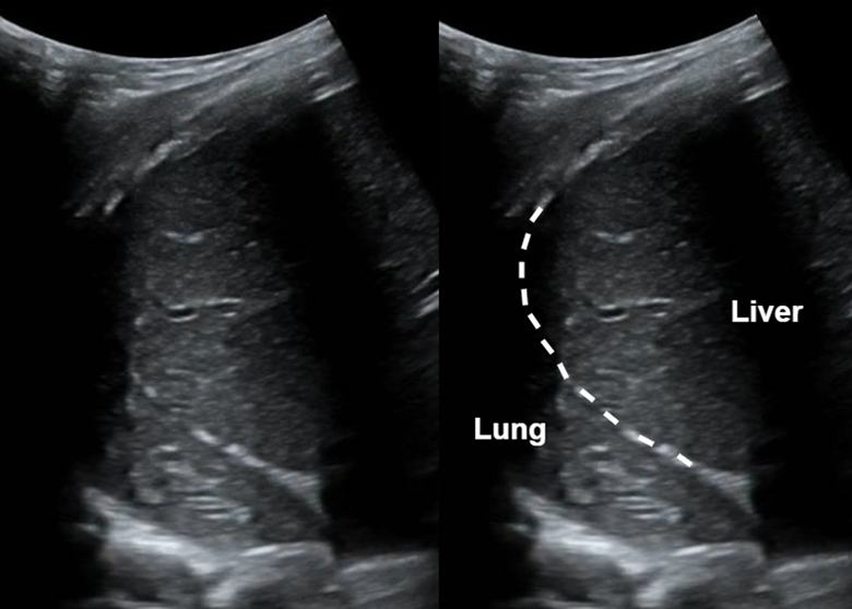
In this LUQ view, we can see free fluid between the diaphragm and the spleen. The diaphragm is highlighted with the dotted white line. Also note the abnormal spleen architecture; the spleen appears to have varying echo intensity with irregular borders and a hypoechoic rim. See the attached clip to see this in real-time!

In this sagittal view of the bladder, we can see free fluid between the bowel and bladder. See the attached clip to see this in real-time!

In this transverse view of the bladder, we can see free fluid above the bladder. See the attached clip to see this in real-time!

Without a doubt this was a positive FAST!

Case Conclusion

The patient was quickly stabilized with IV fluids and pRBC transfusion. CT imaging showed a grade 3 splenic laceration and confirmed moderate amount of hemorrhage in the abdomen and pelvis. The patient was admitted to the PICU for observation.

FAST exam & Splenic laceration

·       With every FAST exam it is extremely important to follow the liver until you have a good view of the liver tip, as fluid will likely collect there first.

·       Remember, free fluid fills potential spaces between structures, so will have characteristically sharp edges! Free fluid will NOT have smooth edges and walls like physiologic structures do.

·       In the LUQ view, any disruption of normal spleen architecture / echotexture could indicate a splenic injury. These include splenic lacerations, hematomas, and rupture.


Happy scanning!

Sono team


Resources to review:

·       https://www.thepocusatlas.com/trauma-atlas

·       https://www.acep.org/sonoguide/basic/fast

·       https://www.ultrasoundcases.info/cases/abdomen-and-retroperitoneum/spleen/trauma-of-the-spleen/

 · 

VOTW: Can’t shrug this one off!

Hello all! This week’s VOTW is brought to you by myself!

Hospital course

An 18 y/o M presented after falling off his skateboard causing him to land on his left shoulder. The XR showed a posterior shoulder dislocation, seen in the XR below where we can see the classic light bulb appearance of the humeral head.

Ultrasound

The left shoulder was scanned with ultrasound, which is shown below.

Note that close proximity of the humeral head to the probe (more superficial), which is indicative of a posterior shoulder dislocation. Also note that the glenoid fossa is not seen well, indicating that the humeral head is not articulating well with the glenoid.

An US-guided interscalene nerve block was completed to relieve the patient’s pain. The target is the brachial plexus cords seen in the image above between the middle and anterior scalene muscles.

In the above clip, we can see the needle entering in-plane lateral to medial with injection of local anesthetic within the interscalene space, surrounding the brachial plexus.

Case Conclusion

After the interscalene block, the patient had great pain control and the shoulder was able to be reduced successfully without any procedural sedation!

This ultrasound of the shoulder was obtained post-reduction. Here we can see that the humeral head is articulating directly with the glenoid.

In this clip, we can see the glenohumeral joint with the humeral head moving well in normal alignment as the shoulder adducts and abducts.


Posterior shoulder dislocation

  • A posterior shoulder ultrasound scan is done by placing the probe just over the scapular spine in a transverse plane, with the probe marker towards the patient’s left.

  • A posterior shoulder dislocation will show the humeral head displaced closer to the probe (appears more superficial on the screen). Also, the humeral head will not be articulated with the glenoid fossa.

  • An US-guided interscalene block can be performed in the ED as a replacement for procedural sedation prior to shoulder reduction as seen with this patient! The interscalene block covers the shoulder and proximal humerus. The probe is placed 2-3 cm superior to the clavicle and the target is the brachial plexus which is commonly referred to as the “stoplight” sign in this view because of its 3 circular hypoechoic structures found between the middle and anterior scalene muscles.


Happy scanning!

Sono team

Resources to review:

 · 

VOTW: “D”-oh! What a heart!

Hello all! This week’s VOTW is brought to you by myself.

Hospital course

30 y/o M presents to the ED after 2 syncopal episodes. He had 10 days of worsening dyspnea on exertion with chest pressure and palpitations. He flew to California 1 month ago and returned yesterday.  

In the parasternal short view of the heart above, we see two cardiac chambers, the right ventricle (RV) and the left ventricle (LV). We can see flattening of the interventricular septum towards the LV chamber, creating a “D”-shaped LV (labelled above).

View the attached clip to see the LV take on a shape of the letter “D” with each contraction! Also note that the RV is dilated and appears larger in size than the ”D”-shaped LV.

The clip above shows a parasternal short view of a normal heart. Notice that the left ventricle appears circular, and the right ventricle forms a smaller crescent-shape surrounding the left ventricle.

Case Conclusion

CT imaging showed pulmonary emboli within the bilateral pulmonary arteries and dilatation of the right atrium and right ventricle associated with right heart strain.

Thrombectomy was deferred because patient was hemodynamically stable. He was started on a heparin infusion in the ED and then admitted.   

Right heart strain

·       Pulmonary embolism can cause an acute increase in pulmonary pressures and right ventricular afterload that causes increased right heart strain. Focused ultrasound has been shown to be both highly sensitive and highly specific in detecting anatomical changes of the heart seen with right heart strain.

·       “D-sign” is a finding suggestive of right heart strain seen on the parasternal view of the heart. Increased right ventricular pressures cause bowing of the interventricular septum into the LV, causing the “D”-shaped LV to form.

·       Other findings suggestive of right heart strain include increased RV size, McConnell’s sign, and TAPSE.


Happy scanning!

Sono team

PS: just in case nobody gets it, the subject line pun is a Simpsons reference!


Resources to review:

·       https://www.thepocusatlas.com/right-ventricular-dysfunction/

·       https://everydayultrasound.com/blog/category/Right+Ventricular+Strain

·       https://www.acep.org/sonoguide/basic/cardiac

 ·