EMS Protocol of the Week - Obstetric Complications

Short cooldown email for today’s EMS PotW (EMS-PoW? Is that catchier?). Protocol 540 – Obstetric Complications, attached below, shockingly addresses potential complications with the obstetric patient. For context, NYC REMAC is using a 160/110 cutoff for severe preeclampsia, associated with some sort of symptomatology. The protocol opens with reference to BLS procedures, which in this case is essentially just checking ABC’s, calling for ALS if needed, and considering IVC syndrome, moving the patient into a left lateral position if indicated. At this point, the only SO ALS intervention is IV access and fluids, and ultimately an OLMC call to you fine folks for discuss magnesium administration.

The MCO for magnesium is written as a 2 gram IV dose over 10-20 minutes, with a repeat dose of 2 grams if needed. Seeing as we normally start at 4 grams for our preeclampsia/eclampsia patients, this allows for an early start for the loading dose at a lower rate while getting the patient to the ED for further eval. Generally, I tend to have a low threshold for authorizing the magnesium, assuming the crew paints a picture of a patient who could benefit from it.

www.nycremsco.org and the protocol binder, both there for you when you need them most.

David Eng, MD

Assistant Medical Director, Emergency Medical Services

Attending Physician, Department of Emergency Medicine

Maimonides Medical Center

 · 
Share

POTD: How to measure Hounsfield Units?

Today we will show you how to actually measure HU on our radiology software, Medview/ PACS.

First open the CT and find the slice where you would like to measure the HU of a particular area. In the first image below, we see some free fluid around the liver which we would like to know whether it is blood or ascites.

Follow the steps in the images below and your HU measurement is on the second line. In this case, the free fluid in this patient's abdomen had a HU of 19.5 which is consistent with ascites.

Stay well,

TR Adam

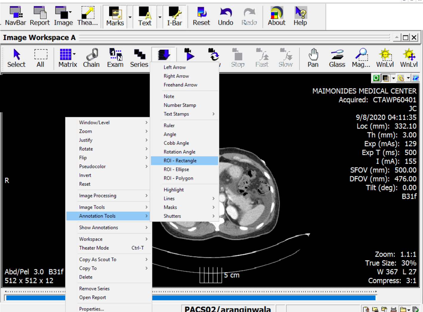
Right click on the image to open the following menu bar

Right click on the image to open the following menu bar

Go to "Annotation Tools" on the menu bar and find and click on "ROI- Rectangle:"


Go to "Annotation Tools" on the menu bar and find and click on "ROI- Rectangle:"

Now create a small box (drag your mouse) over the area you would like the measure the HU. Make sure not to overlap over another area of the slice or it will distort your measurement. You may want to magnify the area you would like to measure

Now create a small box (drag your mouse) over the area you would like the measure the HU. Make sure not to overlap over another area of the slice or it will distort your measurement. You may want to magnify the area you would like to measure

 · 
Share

POTD: Free Fluid in the Belly (Trauma Tuesday!)

This POTD was inspired by a morning report I was present for in Shock Trauma. The case was a 40ish male involved in an MVA. Patient was "shan scanned" for blunt traumatic mechanism. Surgeons saw a ton of free fluid on the scan and took patient directly to the OR. No intra abdominal injuries nor hemoperitoneum were found in the OR. It was later discovered, the patient had a drinking history w/ a cirrhotic liver and the free fluid initially assumed to be blood was actually acetic fluid.

PODT: Free Fluid in the Belly

So you have a trauma patient with a history of alcoholic cirrhosis.  Your FAST is positive but your are unsure whether the free fluid you are seeing is blood from the trauma or the patient's chronic ascites.

You Pan-CT the patient and again all you see is free fluid, unable to tell if what you're seeing is blood or ascites. Your vitals, labs may help guide you. You can also potentially do a diagnosis parenthesis if there is a big enough fluid pocket but this is a very invasive procedure.

What you should do, is look at the Hounsfield Units (HU)!

HU are built into most imaging software and can be used to measure the radiodensity of the material on CT to help distinguish various structures.

Uncoagulated blood typically measures 30 to 45 HU

Clotted blood measures higher at 60 to 100 HU

Ascites/ Plasma measures around 0 to 20HU

So there you have it. You can use HU while looking at your CT images to help you determine whether your trauma patient is bleeding into their belly or what your seeing is more chronic. 

TR Adam

pastedImage.png
An example of hemoperitoneum

An example of hemoperitoneum

An example of hemoperitoneum

An example of hemoperitoneum

An example of plasma/ ascetic fluid

An example of plasma/ ascetic fluid

An example of plasma/ ascetic fluid

An example of plasma/ ascetic fluid

 · 
Share