The Brazilian Wandering Spider

spiderman.gif

Brazilian Armed Spider (Phoneutria)

A box of bananas were delivered to a pub in Bridgwater, England in 2005. Unbeknownst to the person opening the bananas, an aggressive spider was hiding in the bananas and bit him. Later on it would be identified as the venomous and speedy species Phoneutria fera (Brazilian Huntsmen) from South America.

Our topic today is about the Phoneutria species of spiders. There are 8 species, with only a few being the ones that are primarily interesting for envenomation. They all are recognizable by their defensive posture. They have a leg span of about 6 inches and have light stripes under their legs that are revealed while in the posture. When threatened, they assume a characteristic defensive position by raising their four front legs, displaying their fangs, bristling their leg spines, and moving position to continually face their threat. At this point it would be worth mentioning they can jump over a foot in distance.

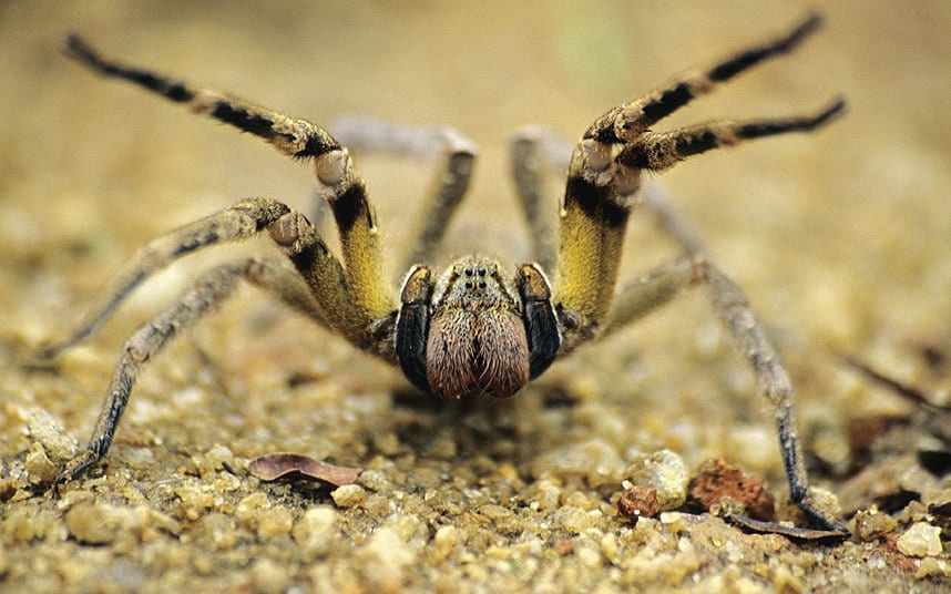
"Come at me bro." - This spider, probably.

Phoneutria fera/Phoneutria nigriventer are the most venemous. P. fera is only in the Amazon while P. nigriventer is in Brazil, northern Argentina, and Uruguay. They are generally only found in the Amazon. Phoneutria nigriventer is also known as a Brazilian armed spider (arantia armadeira in Portuguese).

In their native Amazon, they wander the jungle floor at night and hide inside termite mounds/fallen logs/banana plants. In populated ares they also like to hide in houses/cars and can even be inside boots.

Life Lesson: Always shake your clothes and boots out before putting them on in a tropical country. Keeping your luggage closed is also ideal unless you want a hitchhiker.

Their location spans forests from Costa Rica and Panama down to the Amazon. In South America, this includes Colombia, Venezuela, Guianas, Ecuador, Peru, Bolivia, Brazil, and Paraguay. The importance here is that their location also can include other countries as they sometimes get transported with bananas – 7 of 135 spiders found in north American banana shipments were Phoneutria in 2014.

Toxin: PhTx3 (broad spectrum calcium channel blocker that inhibits glutamate release, calcium uptake, and glutamate uptake in neural synapses). They are aggressive but the good news is that most of their bites do not contain significant amounts of venom. Only 1% of bites result in severe envenomation. Significant envenomation produces severe pain locally followed by both sympathetic and parasympathetic stimulation. This results in a combination of tachycardia/hypertension with nausea, vomiting, diaphoresis, salivation. The most interesting neurotoxic effects are from spinal cord impairment which result in priapism and CNS effects - vertigo, visual changes. Some of the proposed mechanisms for priapism include two chemicals (PhTx 2-5 & PhTx 2-6) which lead to nitric oxide release. Paralysis and pulmonary edema leading to respiratory arrest can occur as well. Children and the elderly are at highest risk for serious envenomation. Most healthy adults recover in 1 to 2 days. Death, shock and ARDS are rare.

Treatment: Local anesthetic infiltration at the bite site can control pain. A polyvalent antivenom (Instituto Butantan, São Paulo, Brazil) is available for cases of severe envenomation from P. nigriventer. If you are far from Brazil, this might be coordinated with your local poison center. Benzodiazpines can help with the excessive tachycardia/hypertension with the goal being symptomatic control with due caution since both sympathetic and parasympathetics are activated. Definite airway should be obtained if significant paralysis. The goal for priapism is detumescence with retention of potency. Aspiration with or without 9% NaCl solution irrigation of the corpora cavernosa may be effective. The current understanding is that priapism is NO mediated so I doubt an α1-adrenergic agonist (100–500 μg/mL phenylephrine solution) instilled into the corpora cavernosa would work. The dosage for states of α1-adrenergic antagonism are 0.5 to 1 mL every 3 to 5 minutes up to 1 hour. Oral terbutaline (5–10 mg) is effective for PGE1-induced prolonged erections but recent studies show the NO effects of the toxin are from a different pathway. If persistent priapism, an operative venous shunt placement may be required. Some studies are being performed with 7-nitroindazole which blocks nNOS (neuronal nitric oxide synthase) to reverse the effects but this is not FDA approved or widely available.

Read More

https://www.researchgate.net/publication/302556178_Phoneutria_nigriventer_Venom_and_Toxins_A_Review

Bucaretchi F, Mello SM, Vieira RJ, Mamoni RL, Blotta MH, Antunes E, et al. Systemic envenomation caused by the wandering spider Phoneutria nigriventer with quantification of circulating venom. Clin Toxicol (Phila) 2008;46(9):885–9. doi: 10.1080/15563650802258524. https://www.tandfonline.com/doi/full/10.1080/15563650802258524?scroll=top&needAccess=true

Bucaretchi  F, Deus Reinaldo  CR, Hyslop  S, et al.: A clinico-epidemiological study of bites by spiders of the genus PhoneutriaRev Inst Med Trop Sao Paulo. 2000; 42: 17. [PubMed: 10742722] 

Vetter RS, Crawford RL, Buckle DJ (2014). "Spiders (Araneae) Found in Bananas and Other International Cargo Submitted to North American Arachnologists for Identification". Journal of Medical Entomology51: 1136–1143. doi:10.1603/me14037

https://www.livescience.com/41591-brazilian-wandering-spiders.html

"Pub chef bitten by deadly spider". BBC News. 2005-04-27


Idiopathic Intracranial Hypertension: Review the essentials of this "can't miss" diagnosis before it's too late!

papilledema.jpg

Idiopathic Intracranial Hypertension (IIH), previously known as ‘Psuedotumor Cerebri’ or ‘Benign Intracranial Hypertension’ that is actually NOT benign is a disorder characterized and defined by the clinical symptoms of increased intracranial pressure (ICP) not due to any other cause. IIH is, in some ways, a diagnosis of exclusion, as you must have negative neuroimaging and a normal CSF composition to rule out other causes of increased ICP prior to making this diagnosis. However, this is not your average “diagnosis of exclusion” since disease progression can lead to significant morbidity.

So, why must you not miss this diagnosis?  Transient vision changes, which is the second most common presenting complaint (after headaches), can lead to permanent vision loss.  And since this is primarily a disease of young, otherwise healthy individuals, you can understand why this is kind of a big deal.

Who gets it?

This is a disorder primarily of overweight females of childbearing age. However, it can affect individuals of all ages and males. Rapid weight gain over a short period of time is potentially a greater risk factor than obesity itself. Classically implicated medications include tetracyclines (doxycycline and minocycline), growth hormone, retinoids and OCPs.

How do these patients present?

The most common complaint: Headache, often diffuse and of gradual onset, which progresses in severity over time. It is frequently accompanied by transient visual changes, which may be precipitated by positional changes (usually standing, sometimes bending forward or lying down), Valsava, eye movement or bright lights. Photopsias (a fancy term for seeing ‘brief sparkles’ or ‘flashing lights’) are frequently described. Pulsatile tinnitus, retrobulbar pain, and back pain round out the list of most frequently seen complaints.

Every complaint listed above is fairly non-specific, so this is where things get a little tricky, right? Keep this in mind: Pulsatile tinnitus (often described as hearing rushing water or wind) in the setting of new headaches is very suggestive of IIH.

Symptoms tend to wax and wane for weeks-to-months, or even years before a diagnosis is made. However, a minority of patients will have a more fulminant course with resulting rapid vision loss.

The most common abnormal findings on exam are papilledema, visual field deficits and sixth nerve palsy.

How is it diagnosed?

You MUST get neuroimaging and send CSF analysis to exclude other causes of increased ICP. And if you are considering IIH, then venous sinus thrombosis should also be on your differential—there is significant overlap in terms of presentation and risk factors.

Once you have excluded all other causes, diagnosis is made via an elevated opening pressure on that LP that you performed (lateral recumbent position).**

**Before the LP, this is the PERFECT opportunity to break out your ultrasound and measure the optic nerve diameter.

In summary, the full list of diagnostic criteria (per the Dandy criteria) are:

  • Symptoms of increased ICP
  • No other neurological abnormalities or impaired level of consciousness
  • Elevated ICP with normal CSF composition
  • Normal neuroimaging
  • No other apparent cause of increased ICP

How is it treated?

For in-service: therapeutic lumbar puncture. Obviously, discontinue any potential offending agents. Weight loss, carbonic anhydrase inhibitors (acetazolamide), diuretics (furosemide) and CSF shunting are all traditionally utilized therapies. Recent data suggests that acetazolamide combined with a low-sodium, weight reduction diet may be the most effective option.

Want to learn more?

https://emedicine.medscape.com/article/1214410-overview

https://www.uptodate.com/contents/idiopathic-intracranial-hypertension-pseudotumor-cerebri-clinical-features-and-diagnosis?search=idiopathic%20intracranial%20hypertension&source=search_result&selectedTitle=1~148&usage_type=default&display_rank=1

https://www.aao.org/eyenet/article/managing-idiopathic-intracranial-hypertension-evid

SaveSave

SaveSave

SaveSave

SaveSave

 · 
Share

POTD: Dog Bites

dog.gif

In celebration of the Year of the Dog, we wanted to cover management of dog bites.

According to CDC data from 2015, there are approximately 4.5 million dog bites per year in the United States with 1 out of 5 requiring medical attention. The wounds tend to be crush injuries with a greater risk of underlying fracture due to the strength of the dog’s jaws. Pay attention to distal neurovascular status, tendon involvement, joint violation and the presence of foreign bodies. A low threshold to x-ray is valuable. These wounds should be debrided and cleaned with well pressured irrigation.

Classically tested, the most common pathogen that creates infection in wounds is Pasteurella Canis (and other Pasteurella species). Immunosuppressed, alcoholics, smokers or asplenic patients should raise concern for Capnocytophagia canimorsus (a gram-negative rod) that causes particularly devastating illness with meningitis and septic shock reported.

Antibiotic prophylaxis/treatment of choice is with amoxicillin-clavulanate 875/125mg twice a day for 10-14 days. Other bacteria of interest include staphylococci, streptococci, and anaerobes. Remember this isn't your run of the mill cellulitis, cephalexin will not cut it.

Repairing these bites has been a subject of debate. REBEL-EM did a great job covering the myths for these wounds with two of the major studies. (link below)

Using 3-7% as a normal wound infection rate for all lacerations, the thought is that you can attempt a closure on some of these wounds for cosmesis. Good indications for closure would be a clean appearing wound that can receive significant irrigation that is <8 hours old. Wounds greater than 8 hours old had greater than a 20% chance of infection if closed in a study by Paschos et al.

Well vascularized areas perform better with closure – the face/scalp. We use non-absorbable sutures and no buried sutures to reduce the burden of foreign bodies present – minimizing infectious risk.

Tetanus should be given to patients suffering dog bites if they have not received it in the past 5 years. Rabies vaccination + rabies immunoglobulin should be considered for dog bites occurring in the USA from dogs that cannot be monitored and/or are unvaccinated. People previously vaccinated against the rabies virus do not need the immunoglobulin but can take part in the 0, 3, 7, 14 series tailored per local infectious disease recommendations.

Dog bites that return to the ED with infection should be cultured (with peripheral smear added for patients at risk of Capnocytophagia). The area should be imaged to assess the integrity of the bone.

Happy year of the dog!

Read More

Centers for Disease Control and Prevention. Preventing Dog Bites. http://www.cdc.gov/features/dog-bite-prevention/index.html. May 18, 2015.

Paschos NK et al. Primary closure versus non-closure of dog bite wounds. A radomised controlled trial. Injury 2014 45(1): 237-40PubMed ID: 23916901

Medeiros IM, Saconato H. Antibiotic prophylaxis for mammalian bite (Review). Cochrane Database of Systematic Reviews 2008 (3); PubMed ID: 11406003

Butler T et al. Capnocytophaga canimorsus: an emerging cause of sepsis, meningitis, and post-splenectomy infection after dog bites. Eur J Clin Microbiol Infect Dis. 2015 34(7): 1271-80. PubMed ID: 25828064

http://rebelem.com/myths-management-dog-bites/