VOTW: In the Thick of It

 ·   · 

HPI

A 40-year-old female with a PMH of polycystic kidney disease, HLD, and HTN presents with 1 month of episodic dizziness. She was referred to the ED by her cardiologist for an abnormal EKG, and had previously been told that she had an enlarged heart. 

Her vital signs are unremarkable. Physical exam is notable for a harsh, blowing systolic murmur. Chest X-ray shows cardiomegaly.

Ultrasound Findings

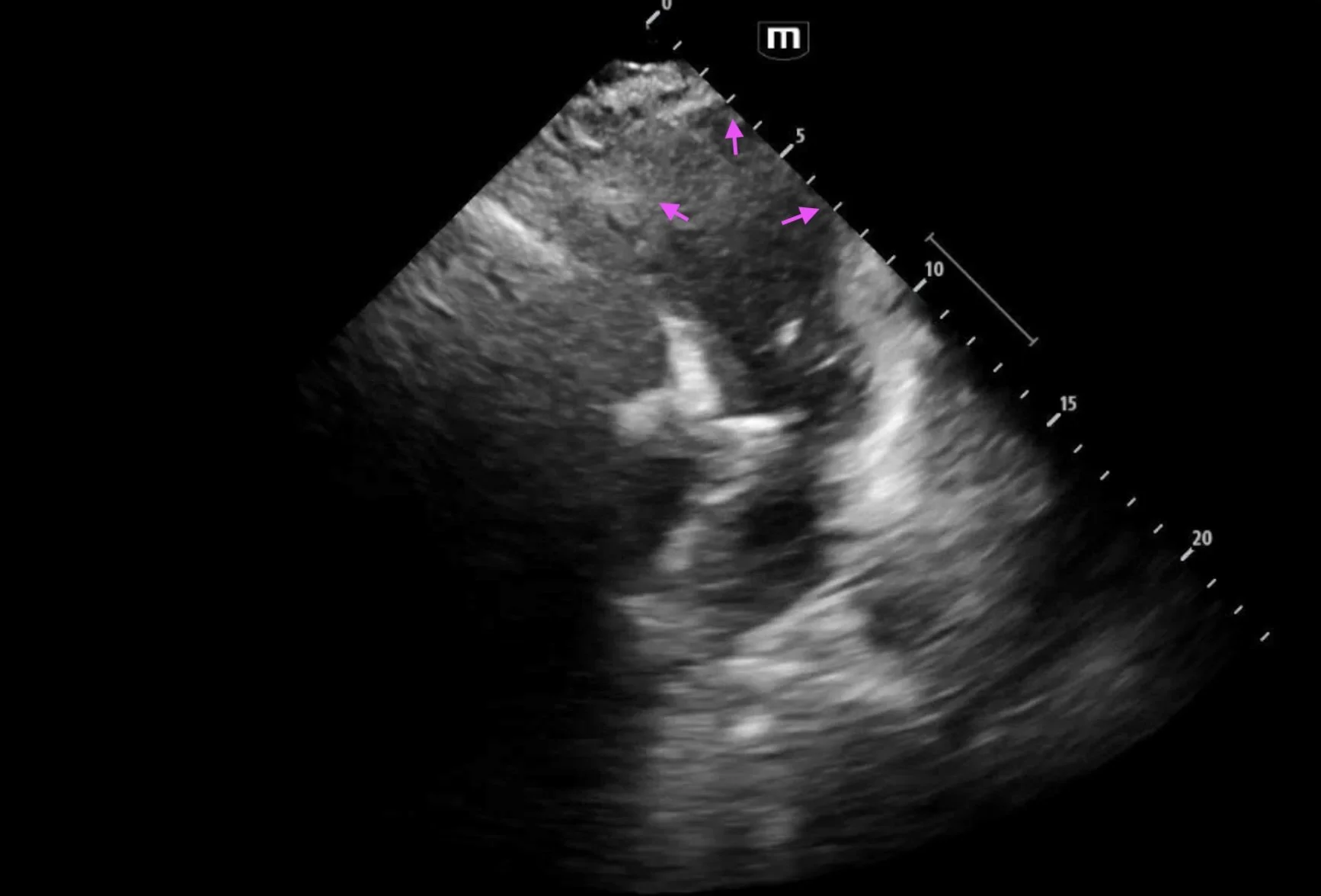
Point-of-care echocardiogram showed no pericardial effusion and was notable for septal thickening to 2.2 cm, concerning for hypertrophic obstructive cardiomyopathy (HOCM).

Echocardiography is the first-line imaging modality for the diagnosis of hypertrophic cardiomyopathy. 

Key findings are wall thickening and intraventricular obstruction. 

  • Wall thickening > 15 mm (or > 13 mm in patients with relatives diagnosed with HOCM). This can be measured in the parasternal long or short axis views. 

  • Interventricular septum to posterior wall thickness ratio of > 1.3 in normotensive patients or > 1.5 in patients with HTN

  • Thickening usually occurs on a focal region of the LV wall

Other associated findings include mitral valve abnormalities, systolic dysfunction, and diastolic dysfunction.

  • Systolic anterior motion of the mitral valve may occur in HOCM due to the Venturi effect. Septal hypertrophy narrows the LVOT, accelerating blood flow and creating a suction force that pulls the mitral valve leaflet into the LVOT. This causes outflow obstruction as well as mitral regurgitation. 

Case Conclusion

Based on these findings, the patient was placed in observation for cardiology evaluation. 

Comprehensive echocardiogram revealed findings consistent with HOCM, including severe asymmetric left ventricular hypertrophy, hyperdynamic LV systolic function (LVEF 76-80%), moderate (grade 2) LV diastolic dysfunction, LV outflow tract obstruction, moderate systolic anterior motion of the anterior leaflet of the mitral valve, and moderate mitral valve regurgitation.

The patient was newly diagnosed with and educated about HOCM. She was discharged with metoprolol 25 mg daily and is anticipated to undergo further treatment with mavacamten and possible septal reduction surgery. 

References & Further Reading

Happy scanning! 


VOTW: Love Trap - A Case of Takotsubo Cardiomyopathy

HPI

A 74-year-old female with a PMH of HTN, scoliosis, and stroke presents to the ED for worsening generalized weakness and poor appetite for 5 days. The patient is tachycardic, tachypneic, and hypoxic to 88%. Her most recent echocardiogram less than a year ago showed a LVEF of 51-55%.

Ultrasound Findings

Bedside echocardiogram showed abnormal wall motion, specifically apical ballooning of the left ventricle

The diagnosis of Takotsubo cardiomyopathy relies on two main criteria:

1) Transient left ventricular wall motion abnormality

2) The absence of a condition obviously explaining this wall motion abnormality

The classic pattern on ultrasound is akinesis of the apex accompanied by hypercontractility of the base, causing an appearance of systolic “ballooning” of the apex. This occurs in 80% of cases; however other variants exist.

Regional wall motion abnormalities extend beyond the distribution of any single coronary artery, sometimes helping differentiate this condition from MI.

Serial echos may show changes in these abnormalities over time.

LV outflow tract obstruction may complicate this condition, causing hemodynamic collapse.

Case Conclusion

ED workup revealed an elevated troponin and BNP, influenza B positive, and the patient was admitted given concern for myocarditis versus ACS.

A comprehensive echo was performed, showing a LVEF of 21-25% and findings consistent with Takotsubo cardiomyopathy. A CTA coronary scan was also performed, showing no evidence of significant CAD. The patient had a complicated hospital course but was medically optimized and ultimately discharged.

References


VOTW: Seeing Things from a Different Angle

HPI

A 58-year-old male presents with decreased vision in the left eye for 4 days. He says he is only able to see shadows through the left eye. Vital signs are within normal limits. Physical exam reveals a fixed pupil in the left eye with significantly decreased visual acuity.

Ultrasound

POCUS reveals complete retinal detachment and posterior lens dislocation (arrow) in the affected eye.

Scanning technique

  • Use a high-frequency linear probe 

  • Avoid excessive pressure on the eye

  • Do not perform ocular ultrasound if there is suspicion for globe rupture

Lens dislocation on ultrasound

  • Subluxation is characterized by deviation of one side of the lens where it has separated from the iris

  • In complete dislocation, the lens can be found within the posterior chamber or vitreous body moving freely with eye movements

  • Dislocation into the anterior chamber is also possible

  • Ocular ultrasound is highly accurate for diagnosing lens dislocation, with a sensitivity of 96.8% and specificity of 99.4% compared to CT imaging

Case Conclusion

This patient was transferred to Kings County for ophthalmology evaluation. 

References

Happy scanning!