Fragile Skin Tears

Today we are going to try to focus on a practical skill which is increasingly important with our aging population: Fragile Skin Tears. Hemostasis/Pain Control:

  • Pressure

  • Use LET (Lidocaine-Epinephrine-Tetracaine)!

  • Topical TXA

  • Surgicel

Suture Techniques:

lac1.jpg
img2.jpg
  1. Apply a couple deep sutures to appose the wound edges. Then place steri strips across the wound and suture through them with 4.0 nylon sutures. This places tension on the tissue below rather than just on the skin.

  2. Place steri-strips parallel to the wound and suture through the steri strips with 4.0 nylon suture. Similar to approach above, however you are able to visualize the wound edges.

  3. Derma-Bond AND Steri Strips. Perform the above techniques, however derma-bond the edges of the wound, let dry, and place sutures through both the steri strips and derma bond. This will be the effective technique for preventing shearing of extremely fragile skin.

img4.jpg

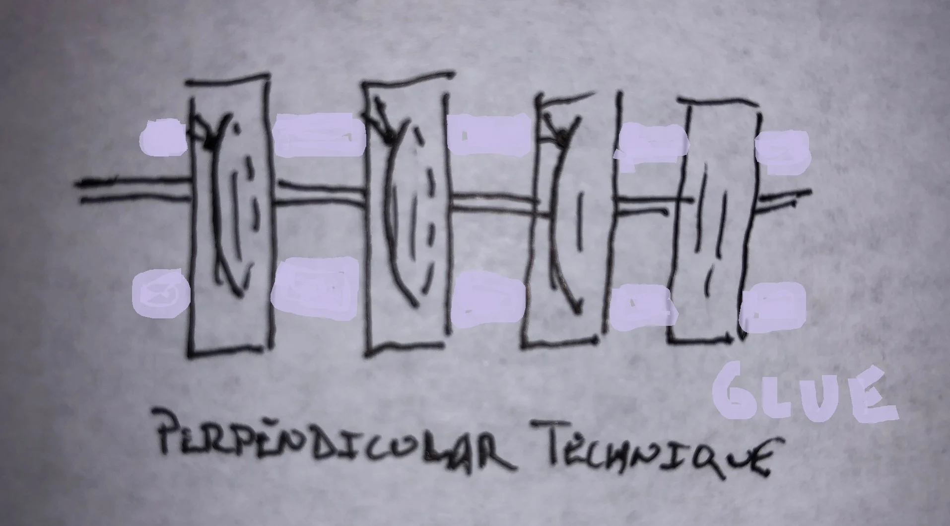
Note there are many variations to this, you may also throw sutures behind the glue. Glue alone may work better for jagged edges than steri-strips. 

  1. Mattress sutures, tegaderm and wait etc.

Aftercare

When the steri strip techniques are used, try to keep wound dry (rather than using topical antibiotics such as bacitracin which will cause the steri strips to become ineffective.  Patients should be vigilant for signs of infection.

Sources:

EMDocs

Lacerationrepair.com

Aliem

Search Terms: Elderly Skin Parchment Laceration Fragile Skin Laceration Tear

 · 
Share

Pearl of the Day: Superior Vena Cava Syndrome

Superior Vena Cava Syndrome Background - effect of elevated venous pressure in upper body that results from obstruction of venous blood flow through SVC - usually caused by external compression of SVC from mass, intravascular thrombosis - most common malignancies: lung, lymphoma - if compression occurs slowly, collateral vessels dilate to compensate for impaired flow - may cause neurological abnormalities from increased intracranial pressure

Signs/Symptoms - usually start 1 - 2 weeks after diagnosis - more common: facial swelling, dyspnea, cough, arm swelling - less common: hoarse voice, syncope, headache, dizziness - rare: visual changes, dizziness, confusion, seizures, obtundation

Diagnosis - chest X-ray -> mediastinal mass - CT chest with IV contrast to assess patency of SVC - MRI if patients cannot receive IV contrast

Management - head elevation to decrease venous pressure - supplemental oxygen to reduce work of breathing - indwelling central venous catheter -> remove - lymphoma suspected -> corticosteroids (very limited evidence in other cases) - cerebral/airway edema present -> loop diuretics, though also very limited evidence - treatment: radiation therapy (can improve symptoms within 3 days), intravascular stents, chemotherapy, catheter-directed fibrinolytics (if secondary to intravascular thrombosis)

Resources Tintinalli's Emergency Medicine, 8th Edition Lepper PM, Ott SR, Hoppe H, et al. Superior Vena Cava Syndrome in Thoracic Malignancies. American Association for Respiratory Care. http://rc.rcjournal.com/content/56/5/653.full. Published May 1, 2011. Accessed April 27, 2018.

 · 
Share

Pearl of the Day: Clavicle Fractures

Clavicle Fractures - clavicle articulates with sternum proximally and acromion distally - protects adjacent lung, brachial plexus, subclavian and brachial blood vessels - mid-portion of clavicle is thinnest and does not contain ligamentous or muscular attachments - signs/symptoms: swelling, deformity, tenderness overlying clavicle; arm is slumped inward and downward; limited ROM at shoulder - diagnosis: usually standard shoulder and clavicle X-rays, but may require 45-degree cephalad tilt view or CT - management: emergent orthopedic consult for open fractures, fractures with neurovascular injuries, fractures with persistent skin tenting

Middle Third Clavicle Fractures - most common - usually managed nonoperatively - risk factors for nonunion: initial shortening > 2 cm, comminuted fracture, displaced fracture > 100%, significant trauma, female, elderly - management: immobilization with either sling or figure-of-eight brace for 4 - 8 weeks - orthopedic follow up in 2 - 3 days: high risk of malunion, severely comminuted or displaced fractures, athletes, professional impact, cosmetic concerns - orthopedic follow up in 1 - 2 weeks for conservative treatment

Distal Clavicle Fractures - type I: fracture is distal to coracoclavicular ligaments with ligaments intact - type II: fracture is distal to coracoclavicular ligaments with disruption of ligaments -> causes upward displacement of proximal aspect of clavicle - type III: intra-articular fractures through acromioclavicular joint - management: types I and III can be managed conservatively with sling immobilization and follow up in 1 - 2 weeks; type II may require operative intervention

Proximal Third Clavicle Fractures - associated with high-mechanism injuries and associated with intrathoracic trauma - diagnosis: CT (also to identify additional injuries) - management: emergent consultation for posteriorly displaced fractures that compromise mediastinal structures; immobilization for all other proximal third fractures - orthopedic follow up in 1 - 2 weeks for conservative treatment

Resources Tintinalli's Emergency Medicine, 8th Edition

 · 
Share