VOTW: Seeing Things from a Different Angle

HPI

A 58-year-old male presents with decreased vision in the left eye for 4 days. He says he is only able to see shadows through the left eye. Vital signs are within normal limits. Physical exam reveals a fixed pupil in the left eye with significantly decreased visual acuity.

Ultrasound

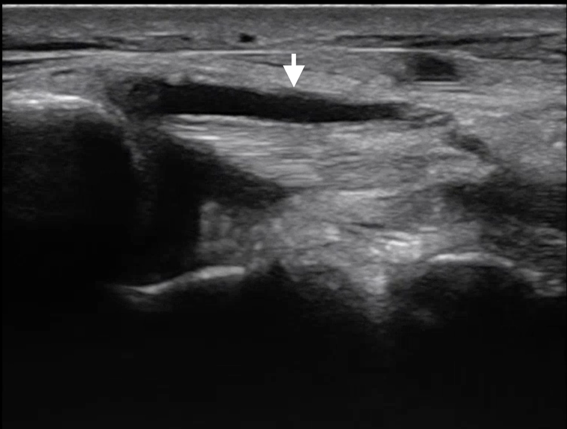
POCUS reveals complete retinal detachment and posterior lens dislocation (arrow) in the affected eye.

Scanning technique

  • Use a high-frequency linear probe 

  • Avoid excessive pressure on the eye

  • Do not perform ocular ultrasound if there is suspicion for globe rupture

Lens dislocation on ultrasound

  • Subluxation is characterized by deviation of one side of the lens where it has separated from the iris

  • In complete dislocation, the lens can be found within the posterior chamber or vitreous body moving freely with eye movements

  • Dislocation into the anterior chamber is also possible

  • Ocular ultrasound is highly accurate for diagnosing lens dislocation, with a sensitivity of 96.8% and specificity of 99.4% compared to CT imaging

Case Conclusion

This patient was transferred to Kings County for ophthalmology evaluation. 

References

Happy scanning! 


VOTW: Tendon to Business

 ·   · 

HPI

A 25M with an unremarkable PMH presents with 3 days of atraumatic left medial ankle pain, redness, and swelling with no other associated symptoms. He is unable to bear weight or ambulate today due to pain.

Physical exam reveals an erythema and tenderness just posterior to the left medial malleolus with no palpable ankle joint effusion.

Ultrasound

POCUS of the left lower extremity demonstrated no DVT in the femoral or popliteal veins, no abscess in the overlying soft tissue, and no joint effusion in the ankle. Fluid was noted surrounding the posterior tibial tendon, shown below in two planes.

The unaffected contralateral tendon is shown for comparison.

Characteristic findings of tendinitis on ultrasound include hypoechoic areas of inflammation, tendon thickening, or peritendinous fluid. POCUS is also useful to assess for other conditions like tendon rupture or tears.

Case Conclusion

CT imaging showed thickening of the posterior tibial tendon with surrounding edema suggestive of tendinopathy with no evidence of abscess. Antibiotics were started for possible infectious etiology. Patient was ambulatory after pain control and was given a referral for orthopedic followup.

Happy scanning!


VOTW: Let It Snow

 ·   · 

HPI

A 20-year-old G2P1 female presents with nausea, vomiting, and back pain x1 week. She is breastfeeding and sexually active. LMP was about 2 months prior, and she had spotting 3 weeks ago. Her vital signs are within normal limits. Patient’s urine HCG in the ED is positive. 

Ultrasound 

Transabdominal ultrasound of the pelvis reveals an irregularly shaped gestational sac with cystic structures, subchorionic hemorrhage, no definitive IUP, and no pelvic free fluid. Transverse (clip 1) and sagittal (clip 2) planes are shown below. 

Case Conclusion

Her serum beta-HCG is found to be >270,000, disproportionate with gestational age. Ultrasound images are consistent with molar pregnancy.  OBGYN was consulted for evaluation, and the patient was scheduled for an outpatient D&C.

Characteristic Findings of Molar Pregnancy on POCUS

  • A complete molar pregnancy appears as a heterogeneous intrauterine mass with many anechoic, fluid-filled sacs, resembling a “snowstorm” or “grape-like” clusters from hydropic swelling of villi. A fetus is absent.

  • Partial molar pregnancies may show abnormal fetal tissue alongside these features.

  • Early detection facilitates timely referral for definitive management, which is important as disease can progress to persistent trophoblastic disease or choriocarcinoma

 Another grape catch on POCUS!

References:


Happy scanning!