VOTW: Ring of Fire

28F G9P1A7 currently 8 weeks pregnant presented to the ED as a transfer from outside hospital for suspected ectopic. Patient reports 6/10 generalized abdominal pain and took Acetaminophen with minimal relief. States that she has been following with outside hospital for her pregnancy and was found to have inappropriately rising HCG levels. Reports vaginal bleeding x3 weeks with irregular menstrual periods. Patient denies fever, chills, nausea, vomiting, chest pain, shortness of breath, dizziness, weakness, calf pain. 

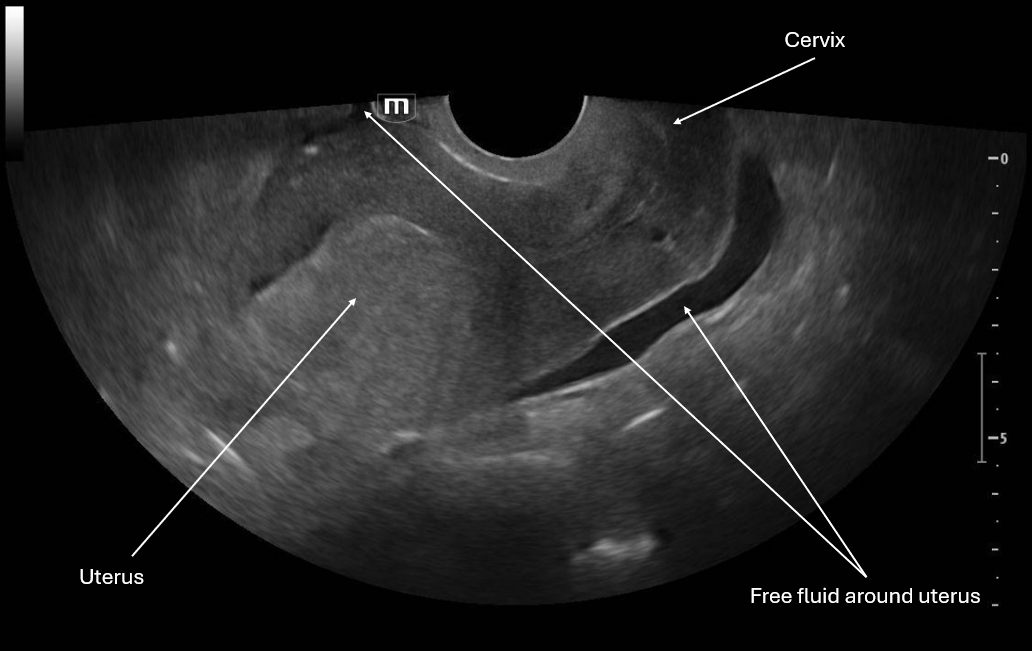
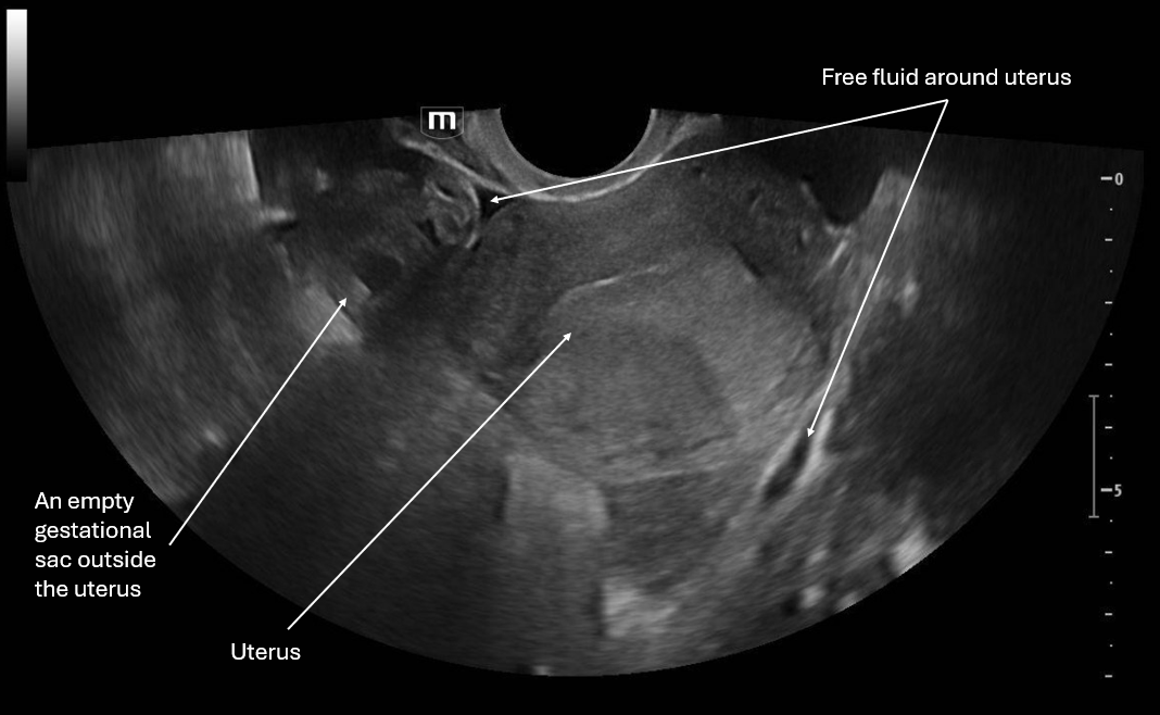
This image is a sagittal view of the uterus with the endocavitary probe marker directed up. From this we can see the fundus of the uterus and cervix with some pelvic free fluid extending past a third of the uterus.

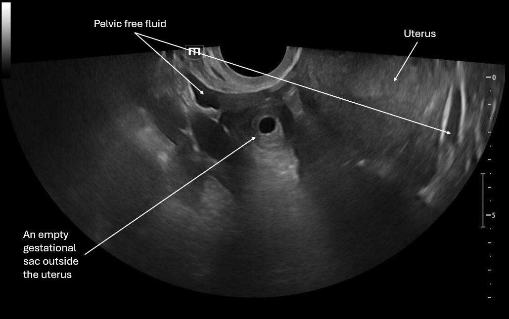
This image is a transverse view of the uterus where the probe marker is towards the patient's right. There is the fundus of the uterus in the middle of the screen and on the patient's right there is what appears to be an empty gestational sac and again we see pelvic free fluid.

This image focuses on what appears to be an empty gestational sac outside the uterus and we again see pelvic free fluid.

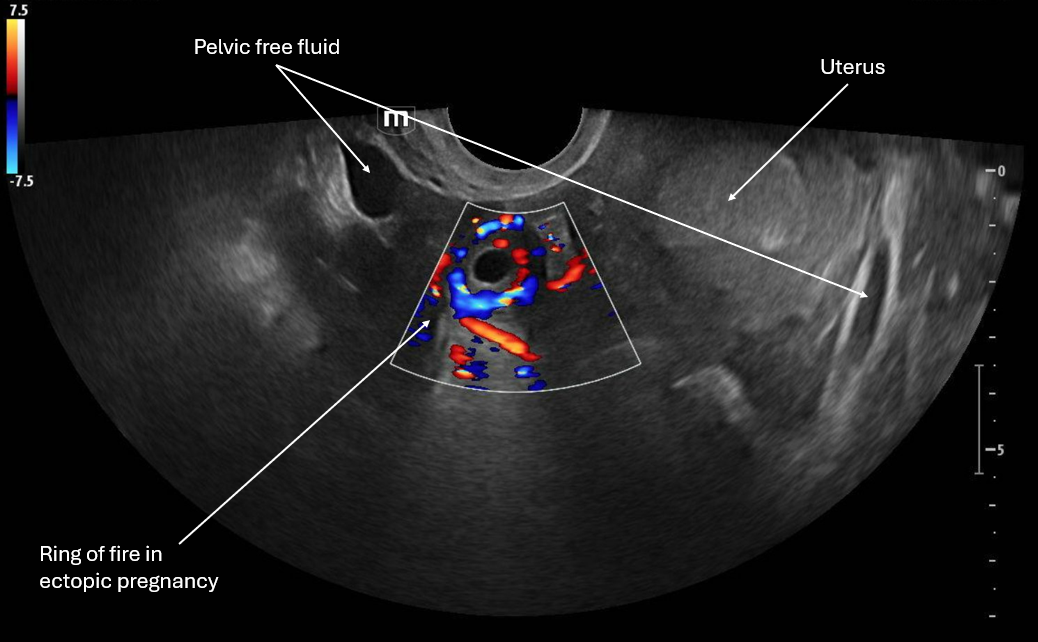
This image applies color flow to the suspected ectopic pregnancy and this shows the "ring of fire" sign which indicates an area of high blood flow due to increased vascularity to the ectopic pregnancy. This "ring of fire" can also be a normal finding with corpus luteum cysts so correlate with other ultrasound findings like free fluid in the pelvis and other elements of the clinical picture.

Patient was taken to the OR and a right laprascopic salpingectomy was performed for a right tubal ectopic pregnancy with about 100cc of blood in the pelvis. Patient was discharged.

POCUS Pearls for Ectopic Pregnancy and "Ring of Fire" sign

  • Circumferential Doppler flow around adnexal mass = hypervascular tissue

  • NOT diagnostic alone (corpus luteum can look identical)

  • Think ectopic if: +hCG + empty uterus + adnexal mass

  • Free fluid in the pelvis, RUQ +/- unstable vital signs is a ruptured ectopic until proven otherwise

 · 

VOTW: Supraclavicular Nerve Block

71 y/o male with PMHx of GERD, presents to the ED via EMS found on floor at bottom of stairwell likely from fall with unknown down time. Patient with bilateral racoon eyes, complaining of right arm pain with right elbow deformity, sling in place. Patient is hard of hearing but patient's family at bedside.

In the first clip we are scanning through to look at the anatomy, this is a sister block to the interscalene and so some of the landmarks are similar with the middle scalene and anterior scalene on either side, but instead of the "stop light" morphology that we see in interscalene block, here we see more of a "bundle of grapes" look. (featuring amazing art from US fellow alumni Dr. Jessie Chen!)

In the 2nd clip we visualize the needle's trajectory as it aims for that supraclavicular bundle. 

Positioning the patient's head to turn to the left as much as possible is important in order to expose the area around the clavicle.

In the 3rd clip we visualize about 10cc of anesthetic being injected around the bundle. Since this was for a quick procedure we used lidocaine 1%. Always remember to calculate the safe amount of anesthetic for each patient you do a nerve block for!

The team was able to reduce the right shoulder successfully!

POCUS Pearls for Supraclavicular Nerve Blocks:

  • Indications include upper extremity fractures (distal to mid-humerus), elbow dislocations, forearm lacerations

  • Use linear probe positioned transverse just above clavicle; head turned away

  • Find artery medial with “bundle of grapes” lateral and visualize the rib deep

  • Rib is the safety backstop and visualize the pleura deep and medial

  • In-plane approach with needle entering lateral towards medial

  • Visualize tip at all times; hydrodissect 1–2 mL before injecting anesthetic

  • Inject 10-15 mL for circumferential spread around the bundle

 · 

VOTW: ESPecial Block

50 y/o male with PMHx of HTN presents to the ED s/p  fall down stairs today. Patient states that he was walking to work then slipped and fell down a whole flight of stairs, striking the L side of his chest with left lateral chest pain. 

Patient had left posterior and anterior rib fractures of ribs 6-10 with a small pneumothorax and some hemothorax, so the decision was made to do an Erector Spinae Plane Block for pain control.

All the clips were recorded with probe marker towards the patient's head and on the left mid-back of the patient's body.

In the first clip we are looking at the landmarks of the ribs and pleura, then tracking more medially and we see the ribs become the transverse processes

Here we are seeing the "flat" areas of the transverse processes and the hazy pleura beneath. This is where we would aim for the needle to go.

In the second and third clip we see the needle trajectory and we see the needle tip at the transverse process and injecting ropivacaine right at the transverse process and lifting the erector spinae muscle off the TP slightly.

The patient reported improvement in pain and was admitted for monitoring due to his injuries.

POCUS Pearls for Erector Spinae Nerve Blocks

  • Use linear or curvilinear probe positioned parasagittal 2–3 cm lateral to midline

  • Transverse process (TP) is the goal landmark and it is a flat hyperechoic line + shadow (NOT rib)

  • Muscle layers include Trapezius to Rhomboid (upper T-spine) to Erector spinae right above the TP

  • Targeting the plane deep to erector spinae / superficial to TP

  • Needle visualization in-plane until TP contact

  • Use 1–3 mL saline to hydrodissect and confirm plane

  • Visualize the fluid spread when the erector spinae “lifts off” TP

  • Inject 20–30 mL (adult) and continue looking for spread

 ·