Not a we problem, an eye problem.

Here is a light visual sampling of eye pathology.

Pterygium:  no acute intervention, follow up pmd +/- opthomology, if impinging on pupil patient will require surgical removal of this benign growth.

Teardop pupil: indicates globe rupture and intraocular foreign body until proven otherwise,  protect globe from any external pressure with eye shield, reduce and prevent IOP elevation- elevate head of bed 30 degrees, analgesia, control hypertension, no valsalva maneuvers, prevent vomiting, EMERGENT optho OR

Hordeolum/Chalazion:  no acute intervention, warm packs, no antibiotics indicated, pcp follow up

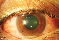
Hyphema: blood in the anterior chamber can be traumatic or spontaneous only needs urgent ophthalmology follow up if up to the pupil. no ED intervention unless signs of intraocular infection then this is an emergency.

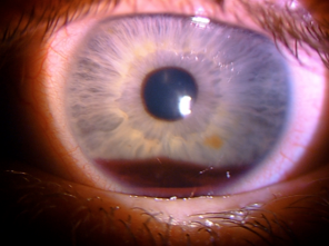
Hypopyon: sign of wide range pathology, can be secondary to trauma, perform woods lamp exam for ulceration, slit lamp exam look for uveitis, urgent opthomology follow up, if infectious etiology suspected systemic antibiotics,

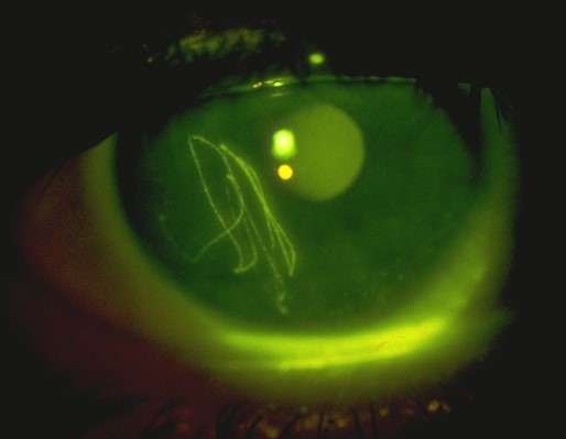
Corneal abrasion: seen on woods lamp exam, raise suspicion for eyelid foreign body be sure to evert both eyelids,  flush eye with saline to remove small foreign body, if abrasion is overlying the pupil antibiotic and urgent ophthalmology follow up.  patient should not wear contacts on injured eye until healed

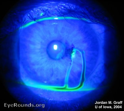
Dedritic ulceration:  Herpetic keratitis treat with po acyclovir or opthalmic antiviral, Urgent opthalmology follow up.

Corneal ulceration: ensure no sign perforation-seidel sign,  opthalmic antibiotics, urgent optho follow up, if patient wears contacts add coverage for Pseudomonas,  do not wear contacts until healed.

Seidel sign: vitreous flowing from site of globe perforation, emergent optho consult in the ER.  protect globe from any external pressure with eye shield, reduce and prevent IOP elevation- elevate head of bed 30 degrees, analgesia, control hypertension, no valsalva maneuvers, prevent vomiting,

Exopthalmos: if in setting of trauma with increased intraocular pressure perform lateral canthotomy,   suspect retrobulbar hematoma or orbital cellulitis,  CT scan emergent optho consult,

Corneal foreign body with rust ring: remove foerign body, urgent follow up for rust ring removal which should be done after 24 hours from initial injury, this is because reepithelialization makes removal easier.

 · 
Share

Do or Di-alysis there is no try... unless you use a trialysis catheter

Ok, lets review the indications for emergent dialysis and the process of placing a dialysis catheter. Indications: -severe acidosis secondary to renal failure or unresponsive to medical therapy -toxic ingestion that is small not protein bound, such as alcohols salicylate, lithium, theophylline, valproate - symptomatic hyperkalemia - symptomatic hypernatremia -fluid overload with oliguria causing respiratory failure -uremia causing encephalopathy, pericarditis, or hemorrhage

placement:

**review your own kit to make sure you have everything you will need and to be mindful of possible extra steps for the following

  1. give anxiolysis if needed

  2. position patient and ultrasound for best place for your catheter

  3. sterilize skin

  4. prep your kit, gown, hat, mask, gloves, sterile cover for ultrasound

  5. flush lumens of your cathetern and apply lumen valves to each lumen but do not place one on the most distal luman through which your wire will pass

  6. have sterile heparin drawn up

  7. drape patient

  8. anesthetize skin

  9. find view use ultrasound guidance and advance needle into vein be sure to be drawing the syringe plunger back to aspirate for blood

  10. once in the lumen advance the needle slightly more in the middle of the lumen to prevent loosing your placement

  11. with non-dominant hand flatten need while making sure to not pull out of the vein

  12. remove syringe from the needle and advance your wire *if there is resistance ultrasound to recheck your placement

  13. advance wire while holding it securely (be sure to always have at least one hand holding the wire)

  14. remove needle

  15. load smaller dilator onto wire

  16. cut the dermis at wire insertion be sure to cutaway from wire

  17. advance dilator and push through skin with twisting motion and inline with trajectory of the wire

  18. remove dilator

  19. load second larger dilator and repeat steps 17 & 18

  20. remove dilator there will be lots of blood good job

  21. advance catheter holding close to the skin with a firm but gentle twisting motion

  22. remove wire

  23. check that all lumens draw back blood with ease

  24. flush each lumen with 1cc of heparin to prevent clotting of the catheter

  25. secure the catheter with sutures

  26. apply sterile dressing

  27. if in internal jugular of subclavian veins confirm placement with xray

 · 
Share

To Ponder Puncturing the Peritoneum?

When and when not to perform paracentesis? Think of the indications for emergent paracentesis in a similar fashion to emergent thoracostomy.  Indications:

  • Relief of respiratory distress caused by massive ascites

  • Diagnostic for infection ie. suspected spontaneous bacterial peritonitis

Contraindications?

  • Overlying cellullitis

  • Vasculature or bowel obstructing desired site

  • Loculated fluid (concern this may be oncologic)

  • Significant coagulopathy INR < 8 platelets >20

2 minute EMRAP video outlining the procedure- https://youtu.be/9npNQM8ANds

Blog post explaining in-series suction canisters-  http://mmcedrco.w02.wh-2.com/EMBlog/suction-cannisters-in-series/


 · 
Share