Dental Trauma

Dental Trauma

dental1.JPG

Pulp – inner layer with neurovascular supply

Dentin – most of tooth under enamel

Enamel – thin outer layer

Cementum – outer layer of root surface

Crown – visible part of tooth

Root – part not visible covered by cementum

Primary/pediatric teeth – 20 teeth, 8 incisors, 4 canines, 8 molars, central incisors start at 7 months of age, 20 by 3 years of age (named A->T)

Permanent Teeth – 32 permanent teeth (8 incisors, 4 canines, 8 premolars, 12 molars), rerupt at 6-7 yo and all 32 by 13 yo

Fracture – evaluate for tooth mobility, sensitivity, fracture

Ellis class I: fracture of enamel, tooth is painless, superficial fracture, no intervention needed, refer to dentistry

el1.JPG

Ellis Class II: dentin fracture, dentin which is has soft golden yellow appearance can be seen, this has higher risk of pulp necrosis/infection, needs f/u with dentistry within 24h, consider dental block for pain relief, CaOH sealant

el2.JPG
caoh.JPG

CaOH paste comes in 2 tubes that need to be mixed then spread over the tooth after it has been dried off (have patient bite down on gauze or soak gauze with epinephrine)

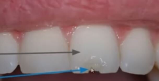
Ellis III: pulp exposure which is a dental emergency, usually very painful unclear neurovascular damage, if pink or bloody discharge at fracture surface, need emergent dental evaluation in ED, high risk of abscess formation

              Cover with CaOH, likely will need root canal by dentistry

el3.JPG

Subluxation – tooth is mobile but not displaced, conservative management w/ soft diet and dental f/u

Luxation – tooth is partially displaced from socket, need tooth splinting for 2-4 weeks, need emergent dental consult

Intrusion – tooth displaced apically, if deep > 3 mm, needs emergent dentistry for repositioning and stabilization, if < 3mm, needs urgent 24h dental f/u

              Consider CTH w/ face for boney fracture, consider CXR for aspiration, XR

Complete Avulsion – loss of entire tooth from socket, dental emergency

1. when did trauma occur – each minute tooth is out reduces viability by 1%, ideally reposition in 15-30 min

2. where is tooth? Aspirated (CXR), swallowed, embedded in oral mucosa, if you have the tooth, touch only the crown, rinse with water and ideally keep the tooth in the socket

              Store tooth in saliva (mouth), milk, saline (less ideal)

3. is tooth primary/permanent

primary tooth should not be re-implanted, needs dental f/u

permanent tooth needs emergent dentistry, periodontal ligament cells can die within 60 min the tooth is outside oral cavity, reimplant tooth to preserve periodontal ligament – splint until definitive management

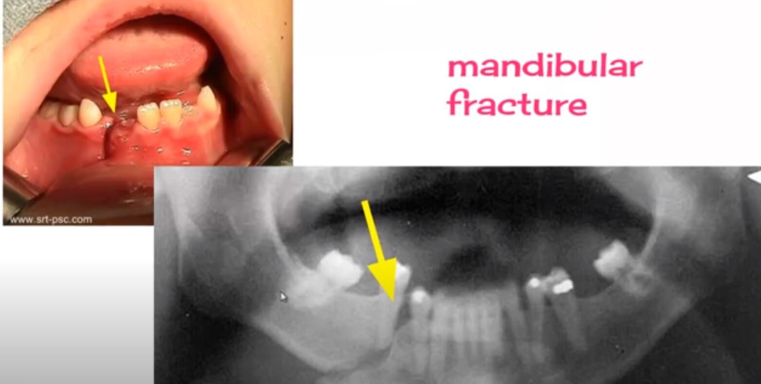
Look for mandibular fracture – can pt open mouth normally, tongue blade test (can pt keep mouth closed to break a tongue blade when twisted), is there pre-auricular tenderness, hematoma at floor of mouth

mendibular.JPG

EMS protocol of the week: Eye Injury

Let’s use this week as a cool down from all the talk of controlled substances over the last couple emails. This will be more of an FYI email, a reminder of what EMS does in the field. Attached are (BLS) Protocol 427 – Eye Injuries and (ALS) Protocol 527 – Chemical Eye Injuries.

427 lays out the initial BLS approach to a patient with an eye injury – they’ll be monitoring ABC’s, assessing for concomitant injuries, provide loose bandaging (WITHOUT putting pressure on the globe) and assisting with contact lens removal, if applicable. If there is concern for a foreign object, they will start irrigation with NS, and if there is an avulsion, they will stabilize, dress, and NOT attempt replacement (presumably after screaming internally, as we all would).

I include the BLS protocol because the ALS protocol starts by referring to BLS procedures (remember 1. that trauma is a BLS call type and 2. that good ALS care starts with good BLS care). The only thing 527 adds is topical analgesia in the form of either Proparacaine or Tetracaine. Did you know that ALS carries eye drops? Crazy!

All of this is Standing Order, so you likely won’t be receiving many OLMC calls with regards to these protocols, but hopefully this helps further your awareness of what sort of care patients may have received in the field.

Reach out with any questions/comments, check out www.nycremsco.org, and don’t forget about the binder!


Diplopia

Diplopia

Case

48F pmh htn, DM presenting with difficulty focusing on distant objects for past few days. Feels like she sees double, all other review of systems normal.  On physical exam, pt has a 6th nerve palsy, but has limited R sided rapid alternating movements. CT/MRI/MAR performed shoulder nonhemorrhagic pontine infarct.

Evaluation

1. Is it monocular diplopia or binocular diplopia? -> have patient cover each eye and see if diplopia persists

If diplopia resolves when either eye is covered -> binocular diplopia

If covering one eye resolves the diplopia -> monocular diplopia in the covered eye

Monocular diplopia – refractive error of eye, retinal disease, less common CNS pathology though possible

              Usually does not require neuroimaging, can send for optho referral

Binocular diplopia – neuromuscular dysfunction causing misalignment of visual fields, CN palsy, oscular muscle dysfunction, brainstem pathlogy

              Usually needs further ED workup for neurologic problem

2. Is diplopia isolated or associated with neurologic signs? Full neuro assessment/exam.

Is there ataxia, vertigo, neuro deficits, dysphagia/dysarthria -> needs rapid neuroimaging and stroke consult to r/o stroke, ideally should have MRI to assess posterior circulation

3. Try to localize lesion casing diplopia especially 3rd and 6th nerve palsies by having patient look horizontally and vertically

3 6 palys.JPG

Isolated nerve palsies are the most common causes of diplopia usually caused by microvascular insult of the extraocular muscles

Isolated 3rd nerve palsy always needs NCHCT and CTA as they are often caused by ischemia or compressive aneurysm

Isolated 4th nerve palsies without other concerning findings can follow with neurology

Isolated 6th nerve palsies usually does not need imaging unless concern for mass/increased ICP, pediatric 6th nerve palsy needs imaging as malignancy is more prevalent than microvascular disease

If there is suspicion of trauma, pt need neuroimaging regardless of whether it’s monocular or not, orbital floor fracture can entrap extraocular muscles or cause a retrobulbar hematoma causing diplopia

Other Diseases to watch for

Cavernous sinus thrombosis – occlusion of cavernous sinus housing CN 3,4,6, often have eye of affected eye, headache, risk factors for thromboembolic disease -> need CT venogram and MRV

Thyroid disease – cause enlargement and fibrosis of extraocular musculature, fatigue, wt loss, TSH/T3/T4

Giant Cell Arteritis - > 50 yo, unilateral HA, ESR/CRP

Myasthenia Gravis – proximal wkns, respiratory distress, fatigable rather than persistent

4. Patients should be dced with neuro/optho f/u as there are many complex motility disorders that need non-emergent MRI or detailed optho exams