EMS Protocol of the Week: Eye Emergencies

Hey all,

This week's protocol looks at eye emergencies that apply to both the adult and pediatric populations.

The prehospital approach starts with CFRs at the most basic level to evaluate and initiate treatments based on these ocular findings:

1) Non-penetrating foreign objects/chemical eye injuries: flush affected eye with NS for 20 minutes

2) Impaled object to eye: use bulky dressings to stabilize object and cover eye to prevent consensual eye movements

3) Avulsed eye: cover eye with saline, sterile dressings and do NOT place eye back into socket

BLS providers provide the additional support of removing contact lenses as needed.

ALS providers provide the additional support of administering proparacaine 0.5% or tetracaine 0.5% drops for chemical eye injuries to assist with irrigation.

Not alot to do on the OLMC side other than to help assist our EMS providers in each ocular scenario.

Check out www.nycremsco.org or the protocol binder on North Side for more.

John Su

PGY-2


Diplopia

Diplopia

Case

48F pmh htn, DM presenting with difficulty focusing on distant objects for past few days. Feels like she sees double, all other review of systems normal.  On physical exam, pt has a 6th nerve palsy, but has limited R sided rapid alternating movements. CT/MRI/MAR performed shoulder nonhemorrhagic pontine infarct.

Evaluation

1. Is it monocular diplopia or binocular diplopia? -> have patient cover each eye and see if diplopia persists

If diplopia resolves when either eye is covered -> binocular diplopia

If covering one eye resolves the diplopia -> monocular diplopia in the covered eye

Monocular diplopia – refractive error of eye, retinal disease, less common CNS pathology though possible

              Usually does not require neuroimaging, can send for optho referral

Binocular diplopia – neuromuscular dysfunction causing misalignment of visual fields, CN palsy, oscular muscle dysfunction, brainstem pathlogy

              Usually needs further ED workup for neurologic problem

2. Is diplopia isolated or associated with neurologic signs? Full neuro assessment/exam.

Is there ataxia, vertigo, neuro deficits, dysphagia/dysarthria -> needs rapid neuroimaging and stroke consult to r/o stroke, ideally should have MRI to assess posterior circulation

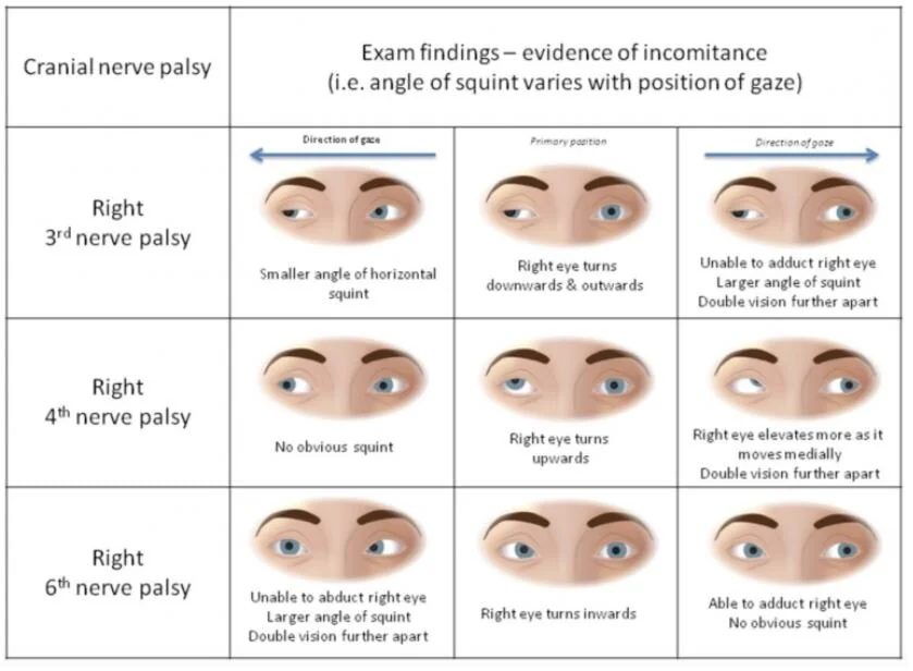
3. Try to localize lesion casing diplopia especially 3rd and 6th nerve palsies by having patient look horizontally and vertically

3 6 palys.JPG

Isolated nerve palsies are the most common causes of diplopia usually caused by microvascular insult of the extraocular muscles

Isolated 3rd nerve palsy always needs NCHCT and CTA as they are often caused by ischemia or compressive aneurysm

Isolated 4th nerve palsies without other concerning findings can follow with neurology

Isolated 6th nerve palsies usually does not need imaging unless concern for mass/increased ICP, pediatric 6th nerve palsy needs imaging as malignancy is more prevalent than microvascular disease

If there is suspicion of trauma, pt need neuroimaging regardless of whether it’s monocular or not, orbital floor fracture can entrap extraocular muscles or cause a retrobulbar hematoma causing diplopia

Other Diseases to watch for

Cavernous sinus thrombosis – occlusion of cavernous sinus housing CN 3,4,6, often have eye of affected eye, headache, risk factors for thromboembolic disease -> need CT venogram and MRV

Thyroid disease – cause enlargement and fibrosis of extraocular musculature, fatigue, wt loss, TSH/T3/T4

Giant Cell Arteritis - > 50 yo, unilateral HA, ESR/CRP

Myasthenia Gravis – proximal wkns, respiratory distress, fatigable rather than persistent

4. Patients should be dced with neuro/optho f/u as there are many complex motility disorders that need non-emergent MRI or detailed optho exams