VOTW: H-appy New Year!

 ·   · 

HPI

29M with no PMH presents to the ED with worsening RLQ pain since last night. He endorses subjective fever, and ROS is otherwise negative. On exam, he has abdominal tenderness to palpation with voluntary guarding.

Bedside ultrasound of the RLQ demonstrates a dilated, tubular, blind-ending structure measuring 17mm. An appendecolith is noted. Findings consistent with acute appendicitis.

CT abdomen/pelvis confirmed the diagnosis of acute appendicitis, with an appendecolith and trace free fluid, for which perforation cannot be excluded.

Pearls

  • For most pediatric patients, choose the linear probe, but can switch to the curvilinear probe for larger patients

  • Place the probe on McBurney’s point or the point of maximal pain and use a lawnmower technique to scan the area

  • Anatomical landmarks: iliac crest, iliac artery, psoas muscle

Signs of appendicitis on ultrasound

  • A non-compressible tubular structure > 6 mm (append-“six") in diameter

  • Sometimes a fecalith (appendecolith) can be seen with posterior shadowing

  • Secondary signs include hyperechoic “hot fat”, free fluid, hyperemia, and bowel wall edema

Case Conclusion

Patient underwent a successful laparoscopic appendectomy. 

Here is a helpful Five Minute Sono video about appendicitis: https://coreultrasound.com/appendicitis/ 

Happy scanning!


VOTW: Tube-y or not Tube-y: Two Cases of Ectopic Pregnancy

Case 1

33-year-old female G3P1, LMP 7 weeks ago, with a history of ectopic pregnancy, which was medically managed, presenting with 1 day of vaginal bleeding. Beta-hCG 5200. 

Transvaginal pelvic ultrasound showed no definitive intrauterine pregnancy and a cystic structure in the left adnexa by the ovary.

In the perinatal unit, ultrasound by MFM confirmed an ectopic pregnancy with a visible fetal heart rate. The patient underwent laparoscopy and salpingectomy that showed a dilated left fallopian tube and had a small amount of intraoperative blood loss. 


Case 2

36-year-old female G4P2, LMP 3 weeks ago, presenting with lower abdominal pain after a bowel movement, followed by vaginal bleeding. Beta-hCG was 230. 

Transvaginal pelvic ultrasound showed no definitive intrauterine pregnancy and a moderate to large amount of free fluid.

The patient underwent diagnostic laparoscopy with salpingectomy. 300 mL of hemoperitoneum was found intraoperatively, and the patient was diagnosed with a left ruptured tubal ectopic pregnancy.

Ectopic pregnancy on ultrasound

Approach

  • Start with the curvilinear probe and switch to the endocavitary probe if better resolution is needed

  • In a patient of childbearing age with abdominal pain and hypotension, start with a FAST exam to look for free fluid in Morison’s pouch

Findings suggestive of ectopic pregnancy

  • Empty uterine cavity or intrauterine fluid without a yolk sac

  • Abdominal free fluid 

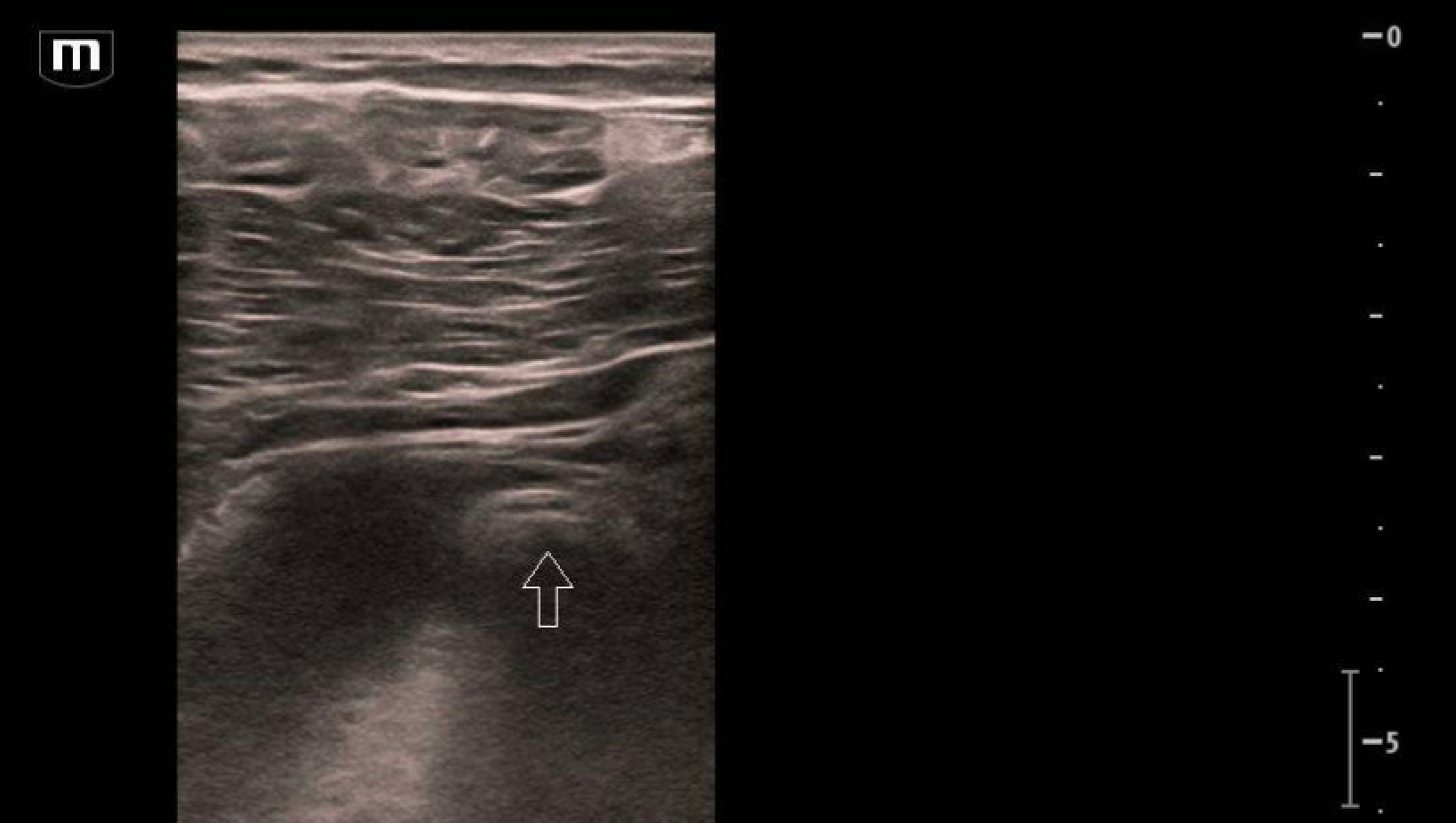
  • A “tubal ring” appearance, an echogenic ring that surrounds an unruptured ectopic pregnancy (n.b. this can be mimicked by a normal corpus luteum). See structure marked by arrow in image above. 

  • Less than 5 mm of myometrium surrounding an eccentrically located gestational sac. This is a type of ectopic pregnancy called an interstitial pregnancy.

References

Happy scanning!

US team


VOTW: A Hairy Situation

HPI: 21 yo male with no PMH presenting for bump noted in the gluteal cleft x 3 days.

The linear probe was placed on the area of interest and showed:

Dot-dash sign/pattern are hyperechoic lines and dots that represent hair. This is commonly seen in ovarian dermoid cysts but also can be seen in pilonidal abscesses from ingrown hair!

Also look carefully at the left side of the screen at the end of the attached video. You might notice the echogenic contents moving around internally as pressure is applied with the probe- this is “squish/swirl sign” AKA “pus-talsis” which is another sign you are looking at an abscess rather than a mass.

As a review of abscesses, you will generally see:

  • A fluid filled irregularly shaped structure with internal septations or echogenic debris (vs a cyst will be contained and completely anechoic)

  • Squish/swirl sign

  • Posterior acoustic enhancement

  • Surrounding tissue cellulitis (early sign: dermal thickening with hyperechoic subcutaneous layer and later sign: “cobblestoning” or edema between fat globules)


Case conclusion: Patient had a bedside I&D of his pilonidal abscess with purulent materials expressed.


Happy scanning!

  • The US Team


Learn more:

  1. https://www.pocus101.com/gynecology-pelvic-ultrasound-made-easy-step-by-step-guide/

  2. https://www.acep.org/sonoguide/procedures/abscess-evaluation

  3. https://coreultrasound.com/cellulitis-vs-abscess/