POTD: C-Collars

Hellooo and welcome to Trauma Tuesday. Today we're going to be discussing c-collars, something that we very frequently see on our patients and very frequently place on our patients.


What is the evidence for it? Are we using c-collars correctly?


C-collars have been recommended by multiple academic societies (surgical, trauma, prehospital, neurological) to be placed pre-hospital if there is a suspected c-spine injury. This recommendation has been in place for ~30 years and has not really changed throughout that time. 


This recommendation has come into question in the past few years given that there aren't many high-quality RCTs truly showing he benefit of c-collars on c-spine injuries and subsequent spinal cord injuries. 


Additionally, conservative estimates show that at least 50-100 patients have c-collars placed on them for every patient that actually has a confirmed c-spine injury - and c-collars are not without harm.


C-collars have been shown to:

  • Increase intracranial pressure via jugular venous compression

  • Increase difficulty for airway management

  • Lead to pressure ulcers when used for an extended period of time

  • Lead to patient discomfort

  • Lead to increased CT imaging that may not have been necessary per our current evidence


Additionally, there is no evidence that small movements of the spine cause worsening c-spine injury. It's large, forceful impacts against the neck that lead to injury, and if the patient has a c-spine injury, they are unlikely to actively move their neck to a degree that will worsen their injury. 


However, given that c-collars are still standard of practice for anyone with a suspected (or confirmed) c-spine injury, we should still follow standard of practice and hospital protocols. Also, it's understandable that we, as EM providers, want to prevent the worst case scenario of a spinal cord injury. 


But I hope this POTD makes us all think harder about how many c-collars we're placing on our patients and the need for better evidence to support (or not support) this practice. 


References

Booth, K, Helman, A. Backboard and Collar Nightmares from Emergency Medicine Update Conference. Emergency Medicine Cases. May, 2015. https://emergencymedicinecases.com/backboard-and-collar-nightmares-emergency-medicine-update-conference/. Accessed October 7, 2024.

Sundstrøm T, Asbjørnsen H, Habiba S, Sunde GA, Wester K. Prehospital use of cervical collars in trauma patients: a critical review. J Neurotrauma. 2014;31(6):531-540. doi:10.1089/neu.2013.3094

Maschmann, C., Jeppesen, E., Rubin, M.A. et al. New clinical guidelines on the spinal stabilisation of adult trauma patients – consensus and evidence based. Scand J Trauma Resusc Emerg Med 27, 77 (2019). https://doi.org/10.1186/s13049-019-0655-x

Plumb, James O.M.Morris, Craig G. et al. Cervical Collars: Probably Useless; Definitely Cause Harm! Journal of Emergency Medicine, Volume 44, Issue 1, e143

https://www.jems.com/patient-care/why-ems-should-limit-use-rigid-cervical/

https://epmonthly.com/article/collar-care/

https://www.emdocs.net/cervical-collars-for-c-spine-trauma-the-facts/

https://www.emra.org/emresident/article/cervical-collar

 · 

VOTW: Mass-ive Fever

This weeks’ VOTW was brought to you by Drs. Hannah Blakely, Patricia Camino, and the ultrasound team that was scanning that day!

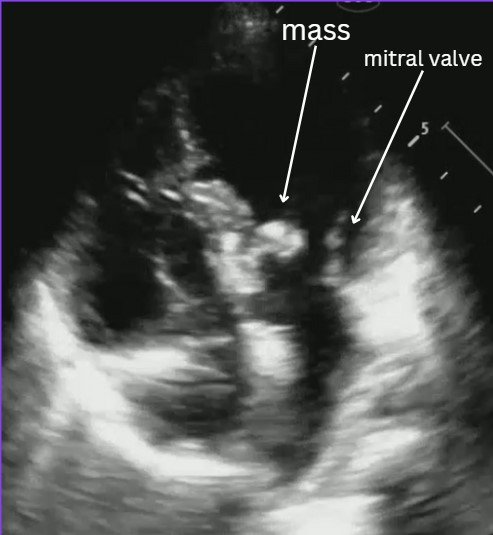
HPI: 6 year old female with PMH of atrioventricular canal defect s/p repair, recent strep throat infection presenting for fever x 14 days.

Bedside POCUS showed:

Evaluating for endocarditis on POCUS:

B mode: look for masses, usually on the lower flow side of the valves (ex: mitral valve- endocarditis is more likely to be found on atrial side).

Color flow: you can usually find associated regurgitation of the affected valve

Possible mimics:

  • Thrombus

  • Papillary muscle rupture/flail leaflet

  • Intracardiac tumor

  • Artifact

Remember that a valve vegetation is one of the major diagnostic criteria for endocarditis. In the right clinical scenario this POCUS finding can highly increase your suspicion for endocarditis.

Case conclusion: CTAP significant for possible splenic and renal infarcts. Patient was admitted for suspected endocarditis. Blood cultures were +MSSA. Pediatric cardiology ECHO was consistent with mitral valve vegetation consistent with endocarditis and septic emboli.

Resources for more info:


Happy scanning!

  • The US Team


VOTW: You take my breath away!

HPI: 90 yo female presenting for worsening shortness of breath and tachycardia x 3 days and right leg pain x 2 weeks with difficulty ambulating.

POCUS showed:

ECHO A4C view (see video): note the size of the RV appears larger than the LV. This is a sign of right heart strain and in the appropriate setting (such as this one) can be concerning for a pulmonary embolism!

Compression views of the common femoral vein (CFV), femoral vein (FV), and popliteal vein (PV). See the echogenic material inside the popliteal vein which is suggestive of a DVT. Remember that during the acute phase of a DVT (<14 days), the clot may appear isoechoic to the blood inside the vein so you may not see this echogenic material and should rely more on your compression exam.

Review on how to do DVT US:

Linear probe

Patient in frog leg position

4 main areas to view

  • Common femoral vein (CFV)-saphenous vein junction (SFV)

    • Clot noted in the SFV within 3 cm to the junction is treated as a DVT. More distally, if there is 5 cm worth of clot noted in the SFV it is also treated as a DVT.

  • CFV branching into [superficial] femoral vein and deep femoral vein

  • Mid/distal femoral vein

  • Popliteal vein

    • Remember the popliteal vein is on top of the popliteal artery (pop on top!)


Tips:

  • You often have to go much higher in the groin than you think to find the CFV-SFV junction

  • Compression testing of the deep veins should not compress the artery (if it is, you’re pressing too hard and can miss subtle DVTs)

  • Deep veins are paired with arteries so identify your landmarks to ensure you are looking at the correct vessels

  • Use your non-scanning hand to help with compression of deeper veins by supporting the other side of the patient’s leg

Case conclusion: Patient with elevated troponin and BNP. CTA significant for bilateral PE’s. Labs and ECHO findings consistent with submassive PE. Patient started on heparin drip and admitted to the floor!

Happy scanning!

  • The US Team