Unstable Pelvic Ring Fractures

The pelvic ring consists of the sacrum and two innominate bones, which are made up of the pubis, ilium, and ischium. These bones are held together by strong ligaments to give the pelvis stability.

A pelvic ring fracture is a severe fracture with 2 breaks in the circular ring, leading to an unstable pelvis and a potentially unstable patient. Fractures that disrupt the pelvic ring predispose patients to bleeding given the large network of arterial and venous anastomoses. Patients who have an isolated pelvic fracture and are hypotensive carry a mortality of 15-40%. Most vascular injuries in the pelvis are venous (90%). While rare, arterial bleeds (10%) should be suspected when a pelvic binder is placed but the patient remains hemodynamically unstable. The retroperitoneal space can accumulate 4 liters of blood before venous tamponade occurs. Pelvic binders are useful in that they can help tamponade bleeding veins, decrease total pelvic volume, and prevent the shifting of bony fragments.

Other unstable pelvic fractures include lateral compression fractures, "open book" pelvic fractures, and vertical shear fractures. Lateral compression fractures occur when a lateral force vector (t-bone in an MVC) causes an anterior ring disruption and sacral fracture.

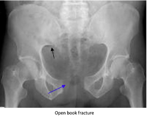
“Open book” fractures occur as a result of anteroposterior compression injury to the pelvis, commonly caused by high-speed trauma or elderly falls. There is a disruption to the pubic symphysis and the pelvis opens like a book. Diastasis of > 1 cm (blue arrow) can indicate instability. Disruption of the pubic symphysis, one of the strongest ligamentous structures in the human body, requires a lot of force and should be a red flag to look for other injuries to the head, spine, chest, or abdomen.

Vertical shear pelvic fractures are seen when one-half of the pelvis shifts upward as a result of a fracture of ipsilateral anterior and posterior pelvic ring fractures. They typically occur as a result of high-energy force applied in the axial direction (aka from the gas pedal to the femur and up to the pelvis). Patients may have an unstable pelvis and leg length discrepancy.

For all unstable fractures, you should appropriately resuscitate and stabilize the patient. Give blood as needed but avoid transfusing through lower limb access because it may drain into the retroperitoneal space. If there is a pelvic ring fracture, consider binding the pelvis. Your binder should lay over the greater trochanters and have enough force to close the pelvic ring (video:https://www.youtube.com/watch?v=tWLBZKeWEkg).


EMS Protocol of the Week: Eye Emergencies

Hey all,

This week's protocol looks at eye emergencies that apply to both the adult and pediatric populations.

The prehospital approach starts with CFRs at the most basic level to evaluate and initiate treatments based on these ocular findings:

1) Non-penetrating foreign objects/chemical eye injuries: flush affected eye with NS for 20 minutes

2) Impaled object to eye: use bulky dressings to stabilize object and cover eye to prevent consensual eye movements

3) Avulsed eye: cover eye with saline, sterile dressings and do NOT place eye back into socket

BLS providers provide the additional support of removing contact lenses as needed.

ALS providers provide the additional support of administering proparacaine 0.5% or tetracaine 0.5% drops for chemical eye injuries to assist with irrigation.

Not alot to do on the OLMC side other than to help assist our EMS providers in each ocular scenario.

Check out www.nycremsco.org or the protocol binder on North Side for more.

John Su

PGY-2


POTD: Intubating the Pregnant Patient

Intubating a pregnant woman is intimidating because you have two patients to consider. Physiologic changes in pregnancy can affect intubation so it is important to plan ahead.


Both ventilation and acid-base status change during pregnancy. As progesterone rises, there is an increase in tidal volume, which results in maternal respiratory alkalosis. This creates a gas gradient to allow for the transfer of CO2 from the fetus to the mom. This maternal hypocarbia causes uteroplacental vasoconstriction, which can cause fetal hypoperfusion and hypoxia. This creates a very delicate acid-base balance that is exacerbated by increased fetal oxygen consumption and CO2 production in the third trimester. In addition, the diaphragm is pushed up by the gravid uterus reducing the mother's functional residual capacity by 10-25%. As a result of these factors, pregnant patients have a shorter safe apnea time and can desaturate quickly.


Progesterone also decreases the tone of the lower esophageal sphincter. Combined with increased intraabdominal pressure from the gravid uterus, pregnant patients are at higher risk for aspiration. For these reasons, you should be careful with bagging and consider intubating in a semi-upright position. This position also has the benefit of taking some pressure off of the patient's chest and IVC.


Anticipate a difficult airway in pregnant patients. Failed intubation is 8x more likely than in the general population. Human placental growth hormone secreted in pregnancy increases blood flow to the upper airways. This results in edema and hyperemia of the airway, causing it to be smaller and more friable. For this reason, you should prepare a smaller caliber ETT. Rocuronium and succinylcholine have been studied with similar efficacy. Induction agents therefore depend on patient specific factors.


TLDR: 1. preoxygenate well due to shorter safe apnea time. 2. Consider a smaller ETT for a narrower and more friable airway. 3. Limit aspiration risks by decreasing bagging if possible 4. consider intubating patients in a semi-upright position.


Thanks for reading! 

Ariella

References: 

https://rebelem.com/respiratory-failure-and-airway-management-in-the-pregnant-patient/

https://www.nuemblog.com/blog/intubating-the-pregnant-patient

https://www.uptodate.com/contents/airway-management-for-the-pregnant-patient