CXR- Consolidation or Atelectasis?

Here is a quick guide on differentiating consolidations vs atelectasis on chest x-ray.

The reason that we can differentiate structures on x-rays is due to differences in density. For example, the lungs are air-filled and appear black whereas the ribs, vertebrae, and heart are solid and appear white. 

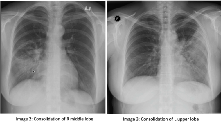
Consolidation: consolidation represents the replacement of alveolar air with fluid, blood, pus, or other substances. There are 3 lobes of the right lung, the upper, middle, and lower lobes. The right middle lobe sits next to the heart border. The left lung has 2 lobes, the upper and lower lobe. The left upper lobe sits next to the heart (image 1). If you have an obscured right heart border, it may indicate consolidation of the right middle lobe (image 2). Similarly, an obscured left heart border may indicate a consolidation in the left upper lobe (image 3). The lower lobes of each lung sit next to the hemidiaphragm. If you cannot make out a hemidiaphragm, it may suggest that there is something of similar density, such as a consolidation, in that lower lobe.

On a normal lateral chest x-ray, the vertebrae should get progressively darker as you get closer to the bases, known as the "more black sign". The vertebrae located near the apex of the lung have overlying muscles, making them appear white, compared to those at the bases that have overlying air, which makes them appear darker (image 4). You should also be able to make out 2 hemidiaphragm on the lateral x-ray with sharp costophrenic angles.

Atelectasis: Atelectasis refers to the collapse of a lung portion. On a normal x-ray, ⅓ of the heart is located on the right and ⅔ of the heart is located on the left side of the chest (image 5). In atelectasis, you will see the mediastinum shift towards the affected side due to volume loss, causing the heart and trachea to shift (image 6). In addition, the unaffected lobe on the ipsilateral side will be hyperlucent as a result of compensatory hyper-expansion. The rib spaces on the affected side may also be closer together when compared to the contralateral side and there may be an elevation of the ipsilateral hemidiaphragm. 

Tip: don’t be fooled by a rotated cxr. Rotation can be assessed by measuring the distance between the medial edges of the clavicles to the vertebral spinous processes. They should be equal or near equal.


Thanks for reading! 

Ariella 

References: 

https://radiopaedia.org/courses/emergency-radiology-course-online/pages/1417

https://radiopaedia.org/articles/lung-atelectasis


US Guided Subclavian Lines

Subclavian lines can be useful when accessing the internal jugular (IJ) or femoral vein is not ideal. For example, you may choose to avoid the IJ in patients who are wearing a c-collar, have a tracheostomy, or have trauma to the head and neck. In addition, some femoral sites are avoided due to pelvic trauma, a large pannus, or a contaminated inguinal region. Using ultrasound to place a subclavian central line can decrease procedure time as well as potential complications, such as pneumothorax, nerve injury, arterial cannulation, thoracic duct injury, hematoma, and hemothorax.

Anatomy: The Subclavian vein is a continuation of the axillary vein as it courses under the clavicle of the first rib and joins with the IJ. The subclavian vein is located inferior and anterior to the subclavian artery. The lung and pleural cavity are deep and interior to the subclavian vein.

There are 2 approaches to placing a subclavian central line: the supraclavicular approach and the infraclavicular approach.

For the supraclavicular approach stand at the head of the bed as if you were placing a central line in the IJ. Place your linear probe above the clavicle to visualize the vein in short axis (you can find the IJ first and trace it until it joins up with the subclavian). Visualize the vein and proceed with Seldinger technique, taking care to note where the pleura is in reference to the vein. You can try to avoid puncturing the pleura by aiming the needle at the rib just under the subclavian vein, so if you overshoot you will be stopped by the rib.

The infraclavicular approach can be done in short or long axis view. If done in long axis, place the probe in the infraclavicular fossa. Your needle in this approach is much more lateral when compared with the landmark-based approach. Identify the point where the axillary and cephalic vein joins with the subclavian and insert your needle past that junction. You will not be able to visualize the artery and vein simultaneously in this view, so use doppler or rotate the probe to short axis so that you can see both the artery and vein at the same time. Proceed with Seldinger technique.

Tip: shrugging the patient’s shoulder opens up a window to puncture the subclavian vein over the 1st rib. In a neutral position, the 1st clavicle lies over the vasculature. Raising the shoulder lifts the clavicle and opens up this space. In this position, if the needle doesn't hit the first rib it will often arch over the first lung, rather than puncture it.

 · 

NG Tube for SBO

Is a nasogastric (NG) tube really needed for management of small bowel obstruction (SBO)? NG tube placement is one of my least favorite ED procedures. I therefore find myself hesitating every time surgery requests one, but what is the evidence behind it?


Surprisingly, there is little data and no randomized control trials on the use of NG tubes in SBO. A chart review in 2013 looked at 290 patients admitted with SBO. 20% of those 290 patients had a NG tube placed. They found that ⅔ of these patients were managed non-operatively, irrespective of NG tube placement. In addition, decompression with an NG tube was not found to be associated with decreased bowel ischemia or need for surgery. Use of an NG tube was actually found to be associated with worse outcomes, such as increased length of hospital stay, higher complication rate, and longer time to resolution.


Part of the reason that I dislike this procedure is the apparent discomfort we cause when placing them. Patients routinely rate it as one of the most painful procedures performed in the ED. We attempt to decrease pain with anesthetics, even though many ED physicians do not believe them to be effective. A RCT was done assessing the use of surgical lubricant versus topical lidocaine and phenylephrine for the nose with tetracaine and benzocaine spray for the throat. Patients reported a significant decrease in discomfort when providers used vasoconstrictors and topical anesthetics compared to surgical lubricant. 


However, there are some cases where NG tubes may be indicated. Patients who are vomiting after antiemetics or have a significantly distended stomach may benefit. Rather than placing them on all patients diagnosed with an SBO, we should select patients for this procedure based on their symptoms.


Thanks for reading!

Ariella 

Resources:

Fonseca AL, Schuster KM, Maung AA, Kaplan LJ, Davis KA. Routine nasogastric decompression in small bowel obstruction: is it really necessary? Am Surg. 2013 Apr;79(4):422-8. PMID: 23574854

Paradis M. Towards evidence-based emergency medicine: Best BETs from the Manchester Royal Infirmary. BET 1: Is routine nasogastric decompression indicated in small bowel occlusion? Emerg Med J. 2014 Mar;31(3):248-9. doi: 10.1136/emermed-2014-203617.1. PMID: 24532357

Singer AJ, Konia N. Comparison of topical anesthetics and vasoconstrictors vs lubricants prior to nasogastric intubation: a randomized, controlled trial. Acad Emerg Med. 1999 Mar;6(3):184-90. doi: 10.1111/j.1553-2712.1999.tb00153.x. PMID: 10192668

Witting MD. "You wanna do what?!" Modern indications for nasogastric intubation. J Emerg Med. 2007 Jul;33(1):61-4. doi: 10.1016/j.jemermed.2007.02.017. Epub 2007 May 30. PMID: 17630077