Warfarin Wednesday

Hey everyone,

After a somewhat heated (well as heated as us third years can get these days) conversation in small group regarding the epistaxis pt with a supra-therapeutic INR I decided to share some info regarding INR reversal based on the current guidelines.
Things to consider:
  • Evidence of active bleeding
  • Magnitude of INR
  • Indicatino for anticoagulation
  • RFs for bleeding
    • Recent bleed w/in 4 wks, surgery w/in 2 weeks, Plt < 50, Liver disease, antiplatelets
  • Volume status?
Options:
  • Vitamin K PO and IV- Warfarin is a VitK antagonist so makes sense right?
    • Similar affects between PO and IV at 24hrs but IV has onset of 6-8hrs
  • FFP- Includes all coagulation factors, has an INR of 1.6
    • VitK dependent factors in concentration of 1U/mL
    • In a 70 kg Patient: 1 Unit Plasma increases most factors ~2.5% 4 Units Plasma increase most factors ~10%
  • PCC (Prothrombin complex concentrate) 3 has Factor 2,9, 10, 4 has 7 also... we only have 4 in our ED I believe.

Bleeding Patient:
  • ALWAYS STOP THE COUMADIN!!
  • INR >1.5 w/ life threatening bleed ( ICH, GI, hemodynamic instability)
    • VitK 5-10mg IV
    • PCC 50IU/kg IV AND FFP 150-300mL
      • If PCC unavailable then 15mL/kg of FFP
  • INR >2 w/ clinically significant but not life threatening bleed
    • VitK 5-10mg IV
    • PCC 35-50 IU/kg IV
  • Minor bleeding:
    • Low risk? Rpt INR next day
    • High Risk or INR >4.5 PO VitK 1-2mg or IV 0.5-1mg and close followup w/in 24hrs

NOT Bleeding:

  • INR <4.5 Omit next dose, resume at lower dose when INR is therapeutic
  • 4.5-10: Omit dose
    • If High risk bleed consider PO VitK 1-2mg or IV 0.5-1mg and pt needs close followup within 24hrs
  • >10: Stop warfarin, VitK, repeat INR at 12-24 hrs
    • High risk patient? Consider PCC 15-30IU/kg
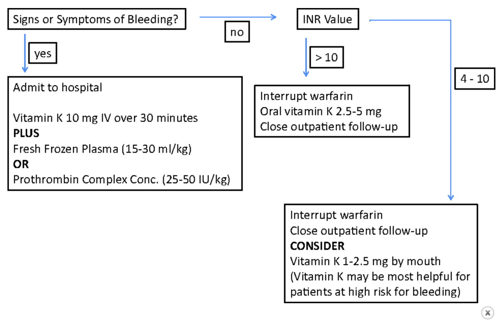
A somewhat simplified algorithm:

A nice concise chart brought to you by our colleagues in Wales:
Sources: Circulation, Surgical Critical Care guidelines, LITFL, CHEST
 · 

Trauma Tuesday: approach to C-spine imaging

For this week's edition of Trauma Tuesday, let's talk about our systematic approach to C-spine imaging in trauma patients, specifically focusing on if/when an MRI is warranted. We all know the initial workup -- Patient arrives in a cervical collar after an MVA. Depending on our level of suspicion for serious c-spine injury based on mechanism and exam, and perhaps depending also on whether the trauma team was called, there are a few ways to proceed: 1) Really low suspicion (minor fender bender, pt was able to ambulate out of the vehicle): apply one of your clinical decision rules (i.e. NEXUS), and if all criteria fit, clear the collar yourself and be done. 2) Moderate suspicion (mechanism still low but pt maaaybe has some midline tenderness on your exam): CT C-spine (has basically replaced x-rays due to superior sensitivity and specificity for detecting C-spine injuries)

Let's say that you proceed with a CT C-spine, and it comes back negative. You go and reassess your patient, who continues to report cervical spine pain upon palpation. What now?

In a large multi-center prospective study published in Dec 2016 by the Western Trauma Association (attached here), the authors concluded that in patients with both a negative CT scan and a normal neurologic exam, the sensitivity was 100% for determining absence of clinically significant C-spine injury.

How did they reach this bold conclusion? Let's backtrack. They included a convenience sample of over 10,000 adult trauma patients who received a CT C-spine after failing NEXUS (breakdown: 45% failed due to distracting injuries, 49% failed due to persistent midline ttp, 5% failed due to abnormal neurologic findings) 90% of these patients ended up with a normal CT C-spine. Of these 90%, a portion ended up getting an MRI C-spine (decision was left up to the treating physician rather than formal protocol). In the end, only 3 patients total were found to have a C-spine injury on MRI when the CT had been negative (in stats-speak, a false negative!) Moreover, all 3 of those patients had an abnormal neurologic exam prompting the decision for MRI, and all 3 were diagnosed with central cord syndrome (classically symptoms worse in upper extremities than lower, often presenting as paresthesias). For CT C-spine imaging alone, this study showed a sensitivity of 98.5% for ruling out clinically significant C-spine injury. Add on the presence of a normal neurological exam, and there were absolutely ZERO patients with significant C-spine injuries who were overlooked with CT C-spine imaging alone.

How's that for evidence? Next time our trauma colleagues might request a knee-jerk MRI C-spine on a patient with iffy "persistent midline tenderness" and a normal CT, we can engage in a respectful and productive discussion about the true utility of such imaging.

TL;DR Take Home Points: Trauma patients who fail NEXUS should get a CT C-spine. CT C-spine alone has excellent sensitivity MRI is indicated in patients with neuro deficits, regardless of negative CT C-spine. Combining your clinical gestalt with evidence-based practice = formula for success.


References: EM:RAP June 2017 https://www.ncbi.nlm.nih.gov/pubmed/27438681

 · 

A "HAT" Trick for Scalp Lacerations!

Welcome to a special short and sweet Christmas Eve edition of POTD!Here's a poem to get us all in the spirit:

Twas the night before Christmas, the ED was quiet, Not a creature was stirring, there wasn’t a riot. The patients slept soundly, so snug in their cots, With some having dreams of free vodka shots...

[Creative credit goes to http://brandtwriting.com/brandts-rants/twas/ where the rest of the poem can be found.]

Okay, down to business. Today we will be discussing some innovative tips/tricks for managing scalp lacerations. Sure, staples and LET are great. But what if you've run out of staple guns, or if it's a scared little kid whimpering in a corner? Is there a less traumatic option?

Hair Apposition Technique - who has heard of this, or better yet, used it on a patient? Apparently it's been around since 2002, but I only learned about this recently!

The Hair Apposition Technique, or HAT trick, creatively uses the patient's own hair essentially as sutures for approximating the scalp lac.

Let me explain the steps:

  1. Irrigate your wound as usual, inspect for foreign bodies
  2. Pull together 3-7 strands of hair on one side of the wound.
  3. Do the same on the other side of the wound.
  4. Twist these two hair bundles in 360-degree revolutions. Do not tie a knot
  5. Secure the intertwined hair bundles by applying a few drops of Dermabond.
  6. Repeat as needed to close the length of the laceration.

For us visual learners, here is an awesome 36 second long video: https://videos.files.wordpress.com/4H47OyPj/hair-apposition-web_hd.mp4

Patient with short hair? Don't worry, you can use the HAT trick, just need 2 pairs of clamps to gain the traction you need:

3 advantages over traditional staples/sutures: 1) Zero pain (especially useful in kids; just tell them you're braiding their hair!) 2) No need to anesthetize the wound (forget waiting for your nurses to first apply LET, then waiting more for it to kick in) 3) No need to return to ED for removal! The hair will unravel on its own after a week.

Bonus: a quick tip for keeping loose hairs out of your field of repair! Next time you're trying to staple Goldberg's latest scalp lac and his hair keeps falling into the lac, try applying petroleum-based ointment (or ultrasound jelly in a pinch) around the area to grease the strands down, then smooth them over to the sides of your field, like so:

This is a super easy/fast way that not only improves your visualization of the lac, but also helps you avoid trapping hair strands within the lac, which could cause wound dehiscense, a foreign body reaction, or a local cellulitis.

Happy lac repairs!

References: https://www.aliem.com/2014/03/trick-trade-parting-hair-scalp-laceration-repair/ https://lacerationrepair.com/techniques/alternative-wound-closure/hair-apposition-technique/

 ·