EMS Protocol of the Week: Eye Emergencies

Hey all,

This week's protocol looks at eye emergencies that apply to both the adult and pediatric populations.

The prehospital approach starts with CFRs at the most basic level to evaluate and initiate treatments based on these ocular findings:

1) Non-penetrating foreign objects/chemical eye injuries: flush affected eye with NS for 20 minutes

2) Impaled object to eye: use bulky dressings to stabilize object and cover eye to prevent consensual eye movements

3) Avulsed eye: cover eye with saline, sterile dressings and do NOT place eye back into socket

BLS providers provide the additional support of removing contact lenses as needed.

ALS providers provide the additional support of administering proparacaine 0.5% or tetracaine 0.5% drops for chemical eye injuries to assist with irrigation.

Not alot to do on the OLMC side other than to help assist our EMS providers in each ocular scenario.

Check out www.nycremsco.org or the protocol binder on North Side for more.

John Su

PGY-2


POTD: Intubating the Pregnant Patient

Intubating a pregnant woman is intimidating because you have two patients to consider. Physiologic changes in pregnancy can affect intubation so it is important to plan ahead.


Both ventilation and acid-base status change during pregnancy. As progesterone rises, there is an increase in tidal volume, which results in maternal respiratory alkalosis. This creates a gas gradient to allow for the transfer of CO2 from the fetus to the mom. This maternal hypocarbia causes uteroplacental vasoconstriction, which can cause fetal hypoperfusion and hypoxia. This creates a very delicate acid-base balance that is exacerbated by increased fetal oxygen consumption and CO2 production in the third trimester. In addition, the diaphragm is pushed up by the gravid uterus reducing the mother's functional residual capacity by 10-25%. As a result of these factors, pregnant patients have a shorter safe apnea time and can desaturate quickly.


Progesterone also decreases the tone of the lower esophageal sphincter. Combined with increased intraabdominal pressure from the gravid uterus, pregnant patients are at higher risk for aspiration. For these reasons, you should be careful with bagging and consider intubating in a semi-upright position. This position also has the benefit of taking some pressure off of the patient's chest and IVC.


Anticipate a difficult airway in pregnant patients. Failed intubation is 8x more likely than in the general population. Human placental growth hormone secreted in pregnancy increases blood flow to the upper airways. This results in edema and hyperemia of the airway, causing it to be smaller and more friable. For this reason, you should prepare a smaller caliber ETT. Rocuronium and succinylcholine have been studied with similar efficacy. Induction agents therefore depend on patient specific factors.


TLDR: 1. preoxygenate well due to shorter safe apnea time. 2. Consider a smaller ETT for a narrower and more friable airway. 3. Limit aspiration risks by decreasing bagging if possible 4. consider intubating patients in a semi-upright position.


Thanks for reading! 

Ariella

References: 

https://rebelem.com/respiratory-failure-and-airway-management-in-the-pregnant-patient/

https://www.nuemblog.com/blog/intubating-the-pregnant-patient

https://www.uptodate.com/contents/airway-management-for-the-pregnant-patient

 · 
Share

CXR- Consolidation or Atelectasis?

Here is a quick guide on differentiating consolidations vs atelectasis on chest x-ray.

The reason that we can differentiate structures on x-rays is due to differences in density. For example, the lungs are air-filled and appear black whereas the ribs, vertebrae, and heart are solid and appear white. 

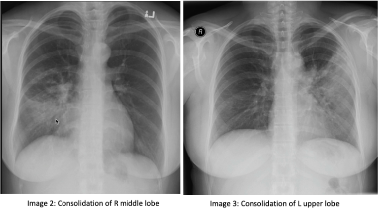
Consolidation: consolidation represents the replacement of alveolar air with fluid, blood, pus, or other substances. There are 3 lobes of the right lung, the upper, middle, and lower lobes. The right middle lobe sits next to the heart border. The left lung has 2 lobes, the upper and lower lobe. The left upper lobe sits next to the heart (image 1). If you have an obscured right heart border, it may indicate consolidation of the right middle lobe (image 2). Similarly, an obscured left heart border may indicate a consolidation in the left upper lobe (image 3). The lower lobes of each lung sit next to the hemidiaphragm. If you cannot make out a hemidiaphragm, it may suggest that there is something of similar density, such as a consolidation, in that lower lobe.

On a normal lateral chest x-ray, the vertebrae should get progressively darker as you get closer to the bases, known as the "more black sign". The vertebrae located near the apex of the lung have overlying muscles, making them appear white, compared to those at the bases that have overlying air, which makes them appear darker (image 4). You should also be able to make out 2 hemidiaphragm on the lateral x-ray with sharp costophrenic angles.

Atelectasis: Atelectasis refers to the collapse of a lung portion. On a normal x-ray, ⅓ of the heart is located on the right and ⅔ of the heart is located on the left side of the chest (image 5). In atelectasis, you will see the mediastinum shift towards the affected side due to volume loss, causing the heart and trachea to shift (image 6). In addition, the unaffected lobe on the ipsilateral side will be hyperlucent as a result of compensatory hyper-expansion. The rib spaces on the affected side may also be closer together when compared to the contralateral side and there may be an elevation of the ipsilateral hemidiaphragm. 

Tip: don’t be fooled by a rotated cxr. Rotation can be assessed by measuring the distance between the medial edges of the clavicles to the vertebral spinous processes. They should be equal or near equal.


Thanks for reading! 

Ariella 

References: 

https://radiopaedia.org/courses/emergency-radiology-course-online/pages/1417

https://radiopaedia.org/articles/lung-atelectasis