Basics of Dermatology 1

POTD: Basics of Dermatology 1

This pearl will be part of a general overview of our favorite subject dermatology. It is very important to have a basic understanding of describing rashes so that our consultants (who will never come to the ED) can understand what we looking at.

(In fact, I believe dermatology only comes once or twice a week inpatient. I admitted a patient for Stevens-Johnson syndrome that didn't see a dermatologist until 3 days after admission)

Story Time – I will be sharing some of my more memorable cases that I’ve seen in relevant POTDs

This case happened during my intern year during my peds rotation.

13 yo female presenting with fever x 2 days with rash. The patient was in the ED 3-4 days ago with similar symptoms and was discharged as herpangina. The patient’s fever was intermittent (not on contiguous days) and subjective, and there were no other associated symptoms. Vital signs stable in the ED; afebrile in the ED. The patient was well appearing tolerating PO.

On her hands and feet were diffuse macular blanching erythematous undemarcated rash that were non-tender. A few of the lesions were patches about 1 x 1 cm. There were some erythematous lesions in the back of her mouth as well. The patient was discharged with a diagnosis of herpangina.

4 days later (third visit), the patient returned again but now with a bit more fatigue and the rash has gotten a bit worse. The patient was still well appearing but the macules have further progressed into patches over the hands and feet. When we re-examined the rash, underneath the blanching patches, there appeared to be non-blanching petechiae underneath. We got basic labs to rule out Kawasaki’s which were all normal and the patient was admitted; other differential included juvenile idiopathic arthritis and lupus.

The patient turned out to have Lupus.

So thinking back, if I had examined her rash more closely, I might have been able to find the petechial rash on the second visit which would have raised my concern for more systemic pathology. My takeaway is to be more thorough looking through all of the areas of rash as well as a benign patch can be covering a more concerning petechiae.

Basics of the Dermatologic exam

Take a history with OLDCARTS as one normally would with any history.

Choose a primary site and describe its morphology, size, shape, dermarcation, color, distribution. Make sure to look throughout the entire body for other lesions. Often times a patient might have an obvious primary lesion and less obvious ones in flexural surfaces can be missed; an isolated rash can be part of a systemic illness. Any associated symptoms like joint pain, abd pain, n/v/d/c, fevers/chills are key as benign appearing rashes can be a part of a more serious systemic illness.

Definitions

Morphology

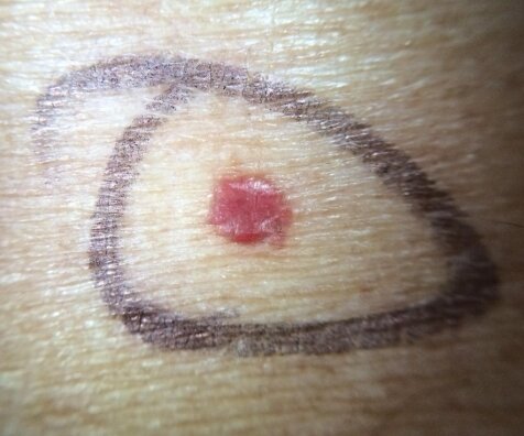
Macule: flat, nonpalpable lesion < 0.5 cm                   Ex: Petechiae is a form of a macule

maculenot pete.jpg

Petechiae – small red or brown macules up to 0.5 cm that don’t blanch with pressure

macule.jpg

Patch: flat, nonpalpable lesion > 0.5 cm                      Ex: vitiligo

vitiligo.jpg

Purpura – circumscribed petechiae more than 0.5 cm in diameter

Purpura_vasculitis_arm_0.jpg

Papule: flat, elevated, palpable lesion < 0.5 cm          Ex: cherry angioma, wart

papule.jpg

Plaque: flat, elevated area > 0.5 cm                              Ex. Psoriasis, seborrheic keratosis

plaque.jpg

Nodule: > 2cm dome shaped with a deep component        Ex. Epidermoid cyst

nodule.png

Vesicle: clear fluid filled lesion < 0.5 cm                                 Ex. Herpes, varicella

vesicle.jpg

Pustule: raised fluid filled lesion < 0.5 cm filled with purulent material (similar to vesicle but with pus)          Ex.  Acne

pustule.jpg

Bulla: clear fluid filled lesion > 0.5 cm                                     Ex. Bullous Pemphigoid, blister

bullous pemphigoid.jpg

Size

Shape

Round/discoid coin shaped, no central clearing (nummular eczema)

Oval ovoid pityriasis rosea Annular round, with active margin and central clearing (tinea corporis) Reticular net-like or lacy (lichen planus)

Linear in a line contact dermatitis Iris/target purple papule in the center of pink macule (erythema multiforme)

Serpiginous snakelike or wavy line track (cutaneous larval migrans)

Polycyclic interlocking or coalesced circles (psoriasis)

Morbilliform measles-like; maculo-papular lesions with confluence on the face and body (roseola, mononucleosis)

Demarcation: are the borders clearly defined? erysipelas vs cellulitis

Color: erythematous (cellulitis, drug reaction), loss of pigmentation (vitiligo), violaceous (Kaposi sarcoma), brown, yellow (xanthoma), black (eschar), honey-colored (impetigo)

Secondary morphology: serum/dried crust (impetigo), fissure, lichenification (thickening of skin), erosion (partial loss of epidermis), ulceration (full thickness loss of epidermis), skin sloughing

Distribution: extensor surface (psoriasis), flexor (atopic dermatitis), generalized (drug eruption), underneath belt/watch (contact dermatitis), dermatomal (varicella)

Dermatology is hard and there is an entire residency for it so we will never know everything about dermatology but there are a few can’t miss dermatologic emergencies that we can’t miss that will be covered in the next POTD!

- Kevin 


Pelvic Binders and Pelvic Fractures

 ·   · 

POTD: Pelvic Binder and Pelvic Fractures

TLDR
If there is pelvic tenderness/instability in trauma with significant mechanism or in an unstable trauma patient, a pelvic binder should be placed to tamponade possible massive hemorrhage and assist in stabilizing fractures.

Please watch these short videos on applying a pelvic binder

https://www.youtube.com/watch?v=8dCntKAExBk        

https://www.youtube.com/watch?v=Omg79Ced6s0      

Today’s pearl of the day covers the use of pelvic binders as well as a general overview of pelvic fractures.

Most pelvic fractures occur due to traumatic, high energy events like a motor vehicle accident. Given its proximity to major organs and blood vessels, they have high morbidity and mortality. (10% mortality for traumatic pelvic fracture, 50% in the unstable patient)

Anatomy

pelvis.jpg

The pelvis is made from the sacrum, coccyx, and the innominate bones (fusion of the ilium, ischium, and pubis). The acetabulum is where the ilium, ischium, and pubic fuse. Pelvic fractures are often classified into acetabular, single bone, and pelvic ring fractures.

Acetabular Fracture

For acetabular fractures, one thing to look for is posterior dislocation of the hip as these need to be reduced within 6 hours of injury. These classically happen when the knee hits the dashboard.

Single Bone Fracture are the most frequently encountered pelvic fractures in the ED and most are stable and are non-operative. For sacral fractures, there is a higher chance for nerve injuries to make sure to complete a full neuro exam.

single.png

Pelvic Ring Fracture

These are the most severe fracture type with two breaks in the circular pelvic ring. They have the highest rate of major hemorrhage.

The Young-Burgess Classification system defines three types of pelvic ring fractures

Lateral compression fracture – most common, T bone MVC

lat.png

Anterior posterior compression fracture – widening of pubic symphysis > 1 cm can represent instability

ap.png

Vertical shear fracture

vert sh.png

Management

If the patient is hemodynamically unstable resuscitate with MTP and give TXA as per normal trauma guidelines

Physical Exam – can rule out significant pelvic injury 93-100% sensitivity in alert patient

Inspect for ecchymosis, deformity, asymmetry, wounds

Palpate for bony tenderness

Compress iliac crest to look for instability; this must be performed gently to minimize hemorrhage if there is a pelvic fracture. Do not rock the pelvis.

If there is a suspected pelvic fracture:

Digital rectal exam for rectal injury i.e. bony fragments, sphincter function, high riding prostate, blood

Genitalia exam – blood at meatus, scrotal, or other hematoma, vaginal exam for vaginal tears

              For urethral injuries a RUG (retrograde urethrogram)

              For bladder rupture a cystogram

Lower limb length discrepancy and malrotation

Neurologic exam


Pelvic Binder

If there is a pelvic ring fracture, a pelvic binder needs to be placed. This is to decrease the space in the pelvis for hemorrhage and stop active bleed. It also assists in stabilizing fractures.

Pelvic binder should ideally be placed prior to intubation as RSI medications can cause expansion of the pelvic space leading to increased bleeding.

This can be with a commercial binder or with a sheet.

https://www.youtube.com/watch?v=8dCntKAExBk         Commercial Binder 78 s

https://www.youtube.com/watch?v=Omg79Ced6s0       Sheet 47s

Studies have shown that sheet binders provide a similar level of stabilization as commercial binders.

bind.png

Often times, there isn’t time for an XR to identify a pelvic ring fracture. If there is instability on physical exam, pelvic tenderness in an unstable patient, or significant mechanism of injury, a pelvic binder should be placed.

Consideration for not applying a pelvic binder: isolated femoral neck fracture, traumatic hips dislocation, foreign object that would be covered up by the binder

Overall, studies have found that pelvic fractures are very safe and have few complications

Imaging options usually starts with an XR and EFAST.

Given that the patient has a pelvic fracture

In the unstable patient with a positive EFAST, the patient should go directly to OR.

In the unstable patient with a negative EFAST, a diagnostic peritoneal lavage should be performed, if positive, the patient should go to OR.

If both EFAST and DPL are negative, a multidisciplinary team of IR, trauma, and orthopedics should be called to assess if the patient needs angiography with embolization, peritoneal packing, or external fixation.

Once the patient is stabilized, a CT abdomen pelvis with IV contrast, and lastly angiography if an active bleed is found.

A great flowchart for Pelvic fractures is shown below.

pelvic-fracture-algorithm-717x1024.jpg

Intrauterine Devices

Studies show that about 10% of US women aged 15-49 use long acting reversible contraception (LARCs) and many of those happen to be intrauterine devices, or IUDs.

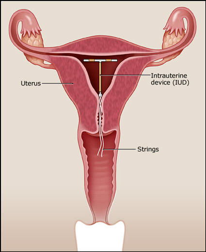
IUDs are a small flexible T-shaped piece of plastic that sit in the uterus and are used for contraception.

iudscehmatic.jpg

What do we as Emergency providers have to know about IUDs and when is it an emergency?

There are 2 categories of IUDs: copper and hormonal.

5 brands are approved for use in the US.

ParaGard (copper)

works by causing an inflammatory response which impairs movement and viability of sperm and diminishes the ability of a fertilized egg to implant

can be used for 10 years

women experience heavy periods/more cramping

string color is clear or white


Mirena, Liletta, Skyla, Kyleena (hormone)

works by releasing a progestin hormone which thickens the cervical mucus, thins uterine wall, and impairs binding of sperm to the egg

can be used for 3-5 years depending on the brand

women experience lighter periods/less cramps

string color is blue or brown


Complications are most commonly see within 1 month of placement and include PID, expulsion, perforation of uterus.


 How to evaluate a patient with an IUD coming in with lower abdominal/pelvic complaints:

 On exam, look for the IUD strings. There are 2 but they can appear as 1. They are generally 2.5cm in length.

 If the strings are too short or not visible at all:

The strings might be curled up in the cervix. Take a cytobrush or Q-tip and sweep the os to see if you can uncurl the strings.

If you still can't see the strings, perform a bedside ultrasound.

If the IUD is in the right location but there are no strings visible, provide outpatient OBGYN follow up.

goodiud.jpg

If the IUD is visible on ultrasound but not in the right location, it might have perforated the uterus. Call OBGYN to evaluate the patient.

If the IUD is not visible on ultrasound, it might have perforated entirely through the uterus. Perform a KUB to make sure it is not somewhere else in the abdomen. If you see it on KUB, again call OBGYN.

If you don't see it on KUB, it's possible that the IUD has fallen out of the uterus. Provide outpatient OBGYN follow up.

kubiud.jpg

If the strings are too long or absent and the IUD is partially out, remove the IUD completely.

If the patient has PID, treat it the same way you would if the patient did not have an IUD and provide OBGYN follow up. Although not removing an IUD in this case goes against logical sense, studies have shown that women who retained their IUDs had similar or better outcomes than women who had their IUDs removed.

Definitely still perform a pregnancy test. There is a 0.1-1.5% risk of pregnancy, depending on which brand of IUD. If the patient is pregnant, be very worried about an ectopic pregnancy and evaluate for it!

If the patient wants to keep her pregnancy and you see an IUP on ultrasound, keep the IUD in and provide OBGYN follow up, assuming patient is otherwise stable and ready to be discharged. Removing the IUD may actually disrupt the pregnancy.

If there is no IUP and patient is stable, asymptomatic, and being discharged, be sure to provide urgent OBGYN follow up.

To remove the IUD, you can use a pair of forceps to pull on the strings. There should be no resistance. If you meet any resistance, stop and call OBGYN.

When removing an IUD, remember to tell your patient that they can become fertile again within a couple days. If they recently had intercourse, sperm can be viable for 5 days, so offer them a Plan B pill.

 · 
Share