VOTW: Distal Radius Fracture

This week’s VOTW is brought to you by myself!


A 72 year old female came in the ED after a FOOSH and suffered a distal radius fracture w/ dorsal angulation seen on x-ray. A POCUS was performed which showed…

Clip 1 shows the dorsal distal radius with sudden cortical disruption and dorsal angulation consistent with the fracture site. The probe marker is facing towards the hand. Clip 2 shows a hematoma block performed w/ ultrasound guidance- the needle is seen entering the fracture site precisely where the fragments meet. Reduction of the fracture was then performed once adequate analgesia was achieved.

Image 1 is prior to reduction. Image 2 is s/p first attempt at reduction. Unhappy with the alignment, reduction was attempted one more time resulting in Image 3 where the alignment is improved. Post-reduction x-rays were obtained, the patient was placed in a sugar-tong splint and discharged with orthopedic follow up.

POCUS for distal radius fractures

In a small study of 83 patients with distal radius fractures, POCUS was 98% sensitive and 98% specific for identifying the fracture when compared to x-rays. Sensitivity and specificity of POCUS ffor the need for reduction was 98% and 100% respectively (1).

While POCUS may not replace x-rays for the management of fractures, it can assist with procedural guidance for hematoma blocks and can evaluate for the adequacy of reduction in real-time rather than waiting for the x-ray tech to come around in between reduction attempts.

How to Identify a fracture

  • Use a linear high frequency probe

  • Visualize the distal radius in its long axis from multiple planes

  • Look for a disruption/angulation in the echogenic cortex

How to perform a ultrasound-guided hematoma block

  • Obtain 10ml of lidocaine drawn up in a syringe, connect it to a saline lock and an injection needle

  • Locate the fracture site using the linear probe

  • Advance the needle into the skin in-line with the probe and guide it into the fracture site

  • Have an assistant inject 10ml of lidocaine into the fracture site

References

Kozaci et al. Evaluation of the effectiveness of bedside point-of-care ultrasound in the diagnosis and management of distal radius fractures. American Journal of Emergency Medicine Volume 33, Issue 1, 2015, Pages 67-71

Happy Scanning!

Your Sono Team


Pelvic Binders and Pelvic Fractures

 ·   · 

POTD: Pelvic Binder and Pelvic Fractures

TLDR
If there is pelvic tenderness/instability in trauma with significant mechanism or in an unstable trauma patient, a pelvic binder should be placed to tamponade possible massive hemorrhage and assist in stabilizing fractures.

Please watch these short videos on applying a pelvic binder

https://www.youtube.com/watch?v=8dCntKAExBk        

https://www.youtube.com/watch?v=Omg79Ced6s0      

Today’s pearl of the day covers the use of pelvic binders as well as a general overview of pelvic fractures.

Most pelvic fractures occur due to traumatic, high energy events like a motor vehicle accident. Given its proximity to major organs and blood vessels, they have high morbidity and mortality. (10% mortality for traumatic pelvic fracture, 50% in the unstable patient)

Anatomy

pelvis.jpg

The pelvis is made from the sacrum, coccyx, and the innominate bones (fusion of the ilium, ischium, and pubis). The acetabulum is where the ilium, ischium, and pubic fuse. Pelvic fractures are often classified into acetabular, single bone, and pelvic ring fractures.

Acetabular Fracture

For acetabular fractures, one thing to look for is posterior dislocation of the hip as these need to be reduced within 6 hours of injury. These classically happen when the knee hits the dashboard.

Single Bone Fracture are the most frequently encountered pelvic fractures in the ED and most are stable and are non-operative. For sacral fractures, there is a higher chance for nerve injuries to make sure to complete a full neuro exam.

single.png

Pelvic Ring Fracture

These are the most severe fracture type with two breaks in the circular pelvic ring. They have the highest rate of major hemorrhage.

The Young-Burgess Classification system defines three types of pelvic ring fractures

Lateral compression fracture – most common, T bone MVC

lat.png

Anterior posterior compression fracture – widening of pubic symphysis > 1 cm can represent instability

ap.png

Vertical shear fracture

vert sh.png

Management

If the patient is hemodynamically unstable resuscitate with MTP and give TXA as per normal trauma guidelines

Physical Exam – can rule out significant pelvic injury 93-100% sensitivity in alert patient

Inspect for ecchymosis, deformity, asymmetry, wounds

Palpate for bony tenderness

Compress iliac crest to look for instability; this must be performed gently to minimize hemorrhage if there is a pelvic fracture. Do not rock the pelvis.

If there is a suspected pelvic fracture:

Digital rectal exam for rectal injury i.e. bony fragments, sphincter function, high riding prostate, blood

Genitalia exam – blood at meatus, scrotal, or other hematoma, vaginal exam for vaginal tears

              For urethral injuries a RUG (retrograde urethrogram)

              For bladder rupture a cystogram

Lower limb length discrepancy and malrotation

Neurologic exam


Pelvic Binder

If there is a pelvic ring fracture, a pelvic binder needs to be placed. This is to decrease the space in the pelvis for hemorrhage and stop active bleed. It also assists in stabilizing fractures.

Pelvic binder should ideally be placed prior to intubation as RSI medications can cause expansion of the pelvic space leading to increased bleeding.

This can be with a commercial binder or with a sheet.

https://www.youtube.com/watch?v=8dCntKAExBk         Commercial Binder 78 s

https://www.youtube.com/watch?v=Omg79Ced6s0       Sheet 47s

Studies have shown that sheet binders provide a similar level of stabilization as commercial binders.

bind.png

Often times, there isn’t time for an XR to identify a pelvic ring fracture. If there is instability on physical exam, pelvic tenderness in an unstable patient, or significant mechanism of injury, a pelvic binder should be placed.

Consideration for not applying a pelvic binder: isolated femoral neck fracture, traumatic hips dislocation, foreign object that would be covered up by the binder

Overall, studies have found that pelvic fractures are very safe and have few complications

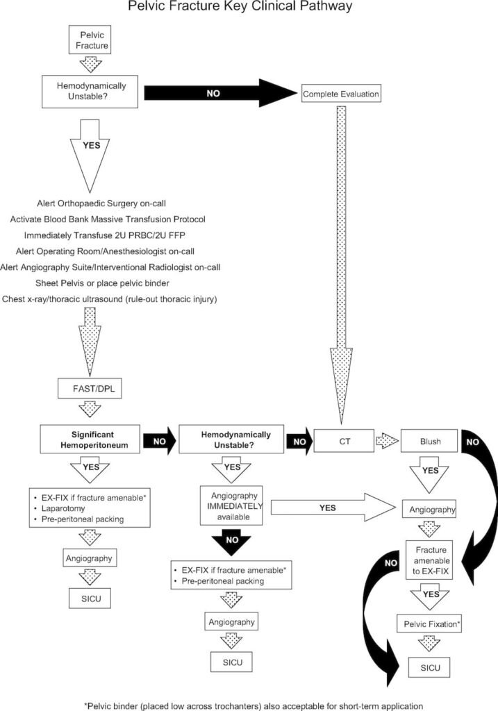
Imaging options usually starts with an XR and EFAST.

Given that the patient has a pelvic fracture

In the unstable patient with a positive EFAST, the patient should go directly to OR.

In the unstable patient with a negative EFAST, a diagnostic peritoneal lavage should be performed, if positive, the patient should go to OR.

If both EFAST and DPL are negative, a multidisciplinary team of IR, trauma, and orthopedics should be called to assess if the patient needs angiography with embolization, peritoneal packing, or external fixation.

Once the patient is stabilized, a CT abdomen pelvis with IV contrast, and lastly angiography if an active bleed is found.

A great flowchart for Pelvic fractures is shown below.

pelvic-fracture-algorithm-717x1024.jpg

5th metatarsal fracture

5MT.jpg

Normal Apophysis in children runs parallel to the bone.  


Dancer’s or Avulsion (pseudo-Jones) Fracture @ cuboid articulation - hard sole shoe for 4-6 weeks and weight bearing as tolerated (WBAT) with orthopedics follow up in a week

Jones Fracture @ intermetatarsal articulation - high risk of non union, pt will need a splint and non weigth bearing activity (NWB) for 6-8 weeks with orthopedics follow up


Metatarsal shaft fracture - high risk of non union, will need a splint, NWB for 10-20 weeks, with orthopedics follow up

Below is a 5 minute video by amazing Dr. Anna Pickens (former Maimo attending) for visual review of the fractures:


http://www.emdocs.net/em-in-5-5th-metatarsal-fractures/

https://youtu.be/4k1dvPdpW4E