TXA in Trauma

Going to take a quick break from the PSA for #traumatuesday

 What is TXA and how does it work?

Tranexamic Acid is a medication used to treat or prevent excessive bleeding.

It works by reversibly binding receptor sites on plasminogen, which reduces conversion of plasminogen to plasmin, further preventing fibrin degradation making up the clot's framework.

txa mechanism.png

Here's a cute animated video showing the mechanism of TXA: https://www.youtube.com/watch?v=emAHFC-Aidg


What role does TXA play in trauma?

CRASH-2 Trial (Clinical Randomization of an Antifibrinolytic in Significant Hemorrhage-2)

  • Double-blinded RCT published in 2010

  • 20,211 patients with traumatic hemorrhage (SBP < 100 and/or HR > 110) or at risk of significant hemorrhage, within 8 hours of injury

  • Dose used was 1g TXA over 10 minutes + 1g over 8 hours

  • All-cause mortality significantly reduced with TXA

  • Risk of death due to bleeding on day of presentation significantly reduced with TXA

  • No significant difference in vascular occlusive events

  • No significant reduction in blood transfusion requirements

  • Greatest benefit seen with early administration (< 1 hour after injury but also < 3 hours). Increased risk of death due to bleeding if administered after 3 hours.


MATTERs (Military Application of TXA in Trauma Emergent Resuscitation)

  • Retrospective observational cohort study published in 2012

  • 896 military personnel who received at least 1 Unit of PRBCs within 24 hours of admission following a combat-related injury

  • Dose used was 1g TXA IV bolus, repeated as deemed necessary by provider

  • All-cause mortality significantly reduced at 48 hours and 30 days especially in patients requiring massive blood transfusion due to their injury


CRASH-3 Trial (Clinical Randomization of an Antifibrinolytic in Significant Hemorrhage-3)

  • Double-blinded RCT published in 2019

  • 12,639 patients with traumatic brain injury (< 3 hours after injury, GCS < 13 or ICH on CT). Patients excluded had major extracranial bleeding, GCS of 3, bilateral unreactive pupils.

  • Dose used was 1g TXA over 10 minutes + 1g over 8 hours

  • Death due to head injury significantly reduced at 24 hours but not at 28 days

  • No significant difference in disability or vascular occlusive events


Bottom Line (based on the literature above):

  • In adult trauma patients in severe hemorrhagic shock for which you are transfusing blood, administer TXA 1g IV over 10 minutes, followed by 1g infused over 8 hours.

  • Reasonable to try TXA for TBI patients with GCS 9-15 and ICH on CT within 3 hours of injury but need more evidence


CRASH-2: https://www.thelancet.com/journals/lancet/article/PIIS0140-6736(10)60835-5/fulltext

MATTERs: https://jamanetwork.com/journals/jamasurgery/fullarticle/1107351

CRASH-3: https://www.thelancet.com/journals/lancet/article/PIIS0140-6736(19)32233-0/fulltext

Reviews from FOAMed sources:

https://emcrit.org/pulmcrit/crash3/

https://www.thebottomline.org.uk/summaries/icm/crash-2/

https://rebelem.com/crash-3-txa-for-ich/

https://first10em.com/the-crash-2-trial/

https://first10em.com/crash-3/

https://emcrit.org/wp-content/uploads/2012/02/TXA-in-trauma-How-should-we-use-it.pdf

 · 

ED Thoracotomy

It’s a new academic year and we are going to start off with some trauma! Today, we are going to talk about ED thoracotomy. It’s basically surgery but in the ED.

First of all, What is ED thoracotomy? It is a Hail Mary attempt in patients with penetrating chest injuries with signs of life in the field and suffer a subsequent witnessed cardiac arrest to open the chest and temporize a life threatening wound. It can be used to relieve cardiac tamponade, control hilar bleeding, cross clamp the descending aorta, or repair a myocardial laceration, all while on the way to the OR for definitive repair.

Success rate and survival is very poor for patients when thoracotomy is performed for blunt trauma, about 2% survival for patients in shock and less than 1% for patients with no vital signs. Conversely, in penetrating wounds, while still poor, 15% of all patients survived.

Guidelines have been established, the so called East and West guidelines, to help balance the already poor odds of survival with the risks of exposing health care providers to blood borne pathogens and salvaging patients with high odds of anoxic brain injury.

Western Trauma Association guidelines: consider ED thoracotomy in patients arriving with blunt trauma and < 10 minutes of CPR or penetrating trauma and < 15 minutes of CPR.

Eastern Association for the Surgery of Trauma: strongest recommendation to perform ED thoracotomy in patients with initial signs of life after penetrating thoracic injury who now present pulseless.

thoracotomy.png

Other commonly quoted numbers. Also consider thoracotomy when you have unresponsive hypotension SBP < 70 despite aggressive resuscitation, chest tube output > 1500cc’s of blood within 24 hours, or when there is > 200/250 cc/hr output of blood after tube thoracostomy for 2-4 consecutive hours.

What you need: A well-staffed and properly trained team that includes also includes a trauma surgeon. Lots of PPE. Thoracotomy tray (rib spreader, #10 or #21 scalpel, scissors, foreceps, vascular clamps, curved artery forceps, needle driver, internal defibrillation paddles, skin stapler, sutures). I would also add a foley catheter as it can be inserted into the heart and the balloon inflated to stabilize a laceration while sutures are being placed.

How is it done? These are the basic steps. Secure the airway and insert an NGT, it can help distinguish the aorta which lies posterior to esophagus. Then, start with a L sided approach, place a chest tube on the R side concurrently. Incise from the sternum to the posterior axillary line at the 4/5th intercostal space, cutting through skin/soft tissue and muscle in one go. Spread the ribs with the rachet bar down. Pick up the pericardium and open it. Inspect myocardium for lacerations. Cardiac massage, internal defibrillation, and intracardiac epinephrine can be done. The aorta can be cross clamped for up to 30 minutes if hypotension still persists. If no evidence of L sided injury, extend to the R side (clam shell).


Hope this helps next time you get a note about a stab or GSW, active CPR, 3 minutes out!

Sources

https://emcrit.org/emcrit/procedure-of-thoracotomy/

https://i0.wp.com/emcrit.org/wp-content/uploads/2012/10/CJLDP_bW8AAVQ2o.png-large.png

https://westerntrauma.org/documents/PublishedAlgorithms/WTACriticalDecisionsResuscitativeThoracotomy.pdf

https://www.east.org/education/practice-management-guidelines/emergency-department-thoracotomy

https://jamanetwork.com/journals/jamasurgery/fullarticle/391389

https://wikem.org/wiki/Thoracotomy

West guidelines algorithm.jpg
 · 

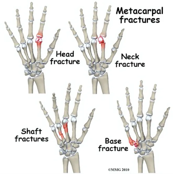
Metacarpal Fractures

Trauma Tuesday!Metacarpal Fractures

mcp fractures.png


Why do we care so much about a few small bones in the hand? Because missed injuries can lead to permanent disabilities--we (as well as our patients) need our hands for pretty much everything.

How to assess for these injuries? Do your typical hand exam but pay special attention to:

Rotational alignment! Have the patient flex at the MCP and PIP, forming a loose fist with the DIPs extended (as in the figure below, to the left). The axis of each digit should merge near mid wrist. Rotational malalignment will cause deviation of this axis for the injured digit.

mcp fractures 2.png


Rotational malalignment is usually an indication for operative repair, so be sure to check for it. 

Don't forget: Any open wound over the MCP should alert you to the possibility of a "fight bite"--usually require exploration or washout. This needs EMERGENT ortho evaluation. 

Diagnosis

Get X-rays - AP, lateral, and oblique views; pay special attention to the lateral as this is what you will use to measure angulation.

For the quick and dirty: acceptable shaft angulation is 40° for 5th MC, 30° for 4th MC, 20° for 3rd MCP, and 10° for 2nd. Reduce if there is greater angulation. 

Management

NONOPERATIVE: For stable fractures, those without rotational deformities, and those with acceptable angulation and shortening (usually 2-5mm for each shaft) => nonoperative repair: 

Reduce a dorsally angulated neck fracture before splinting, usually done via the Jahss technique. (https://youtu.be/40irKoUJqsM)

For MCP head/neck/shaft fractures, radial or ulnar gutter splint depending on which MCP is injured. For MCP base fractures, wrist splint. 


OPERATIVE: For open fractures, intra-articular fractures, fractures with rotational malalignment, significantly displaced or angulated fractures, or in the event of multiple MCP fractures => operative repair

Err on the side of prompt orthopedic follow up. 


Sources

https://emergencymedicinecases.com/episode-29-hand-emergencies/

https://coreem.net/core/metacarpal-fractures/

https://www.orthobullets.com/hand/6037/metacarpal-fractures

 ·