Diplopia

 ·   · 

Where to start?

First, figure out is it monocular or binocular diplopia?

Monocular: diplopia persists with one eye closed

Almost exclusively an eye problem.  It is almost always benign and most often due to refractive error. Give ophthalmology referral and no further imaging is indicated unless warranted by other signs or symptoms.

Binocular: diplopia resolves with one eye closed

Often due to neuromuscular dysfunction causing misalignment of gaze. 

This requires further evaluation in the ED to rule out acute neurological injury.


Next, is the diplopia isolated or associated with other neurologic signs or symptoms?

Do a thorough neurological exam. 

If you find any other neurological symptoms or signs, consider a stroke code/consult and get CT/MRI.  

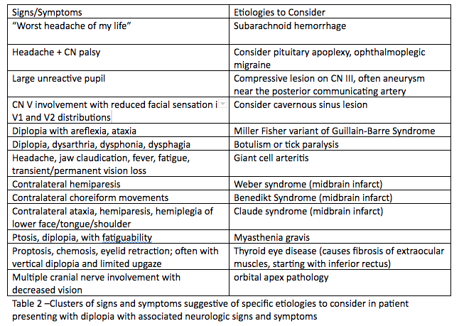
Think of the following table of diagnoses for patients presenting with diplopia and focal neurological deficits, and order the appropriate tests and imaging needed:

Table for Diplopia DDx.png

If there is an isolated palsy, try to localize the lesion.

Ocular Palsies.png

Isolated 3rd nerve palsy: may be due to a cerebral aneurysm, especially if affecting the pupil, and must be emergently evaluated with noncontrast head CT and CTA

Isolated 4th nerve palsy: usually present with vertical or diagonal diplopia, usually due to trauma or idiopathic; be sure to check for cerebellar signs because the 4th nerve exits on the dorsum of the brainstem and can be compressed by a posterior fossa tumor. If no cerebellar signs or trauma, can be discharged with an ophthalmology referral.

Isolated 6th nerve palsy: present with horizontal diplopia, usually idiopathic, check for bilateral 6th nerve palsies as this can be a sign of a tumor (some sources state an isolated 6th nerve palsy should also be evaluated with head imaging prior to dispo). 

For horizontal diplopia, evaluate for internuclear ophthalmoplegia, and if present, get an MRI. Various causes, including multiple sclerosis. 

In children, there is a higher prevalence of malignancy (vs microvascular insult) so they always require head imaging.

Now what?

The need for neuroimaging in diplopia will depend on the palsy the patient has as well as the presence of other abnormalities in the history or physical exam. 

Isolated 4th nerve palsies and 6th nerve palsies can be referred to a specialist for evaluation and do not require imaging in the ED (except as outlined above).

The presence of a 3rd nerve palsy, multiple concomitant palsies, evidence of papilledema, infection, trauma, or cavernous sinus thrombosis require urgent imaging in the ED.

  • 3rd cranial nerve involvement: CTA

  • Increased ICP: CT head

  • Associated neuro deficits/complex motility disorders: CT head, preferably MRI

  • Suspected infection: CT head/orbits with contrast

  • Suspected Cavernous Sinus Thrombosis: CTV, followed by MRV if CTV negative

Patients over age 50 with diplopia should have inflammatory markers ordered.
Diplopia merits urgent referral to an ophthalmologist or neurologist once immediate, intervenable causes have been ruled out.

Sources

http://www.emdocs.net/diplopia-evaluation-and-management/

https://www.nuemblog.com/blog/double-vision

https://crashingpatient.com/wp-content/uploads/2018/06/diplopia.pdf

 · 
Share

Trauma Tuesday: Who is Rolando Bennett?

 ·   · 

What is it?

Bennett fracture: a partial intraarticular fracture of the base of the 1st metacarpal

Bennett.jpg


Rolando fracture: a Y or T shaped complete intraarticular fracture of the base of the 1st metacarpal


Rolando.jpg

Why do we care? 

Could you imagine going through life without working thumbs?

80% of thumb fractures involve the metacarpal base.

Bennett and Rolando fractures are associated with a high risk of early arthritis and mechanical limitations, especially if reduced inappropriately.

When to suspect it?

Usually caused by axial force applied to the flexed thumb.

Patients will experience pain at the base of the thumb, with possible swelling, ecchymosis and tenderness to the area, and worsened pain with range of motion.

For all suspected thumb fractures, get dedicated thumb Xrays, but if you see normal Xrays and still have a high clinical suspicion for injury, get a true AP view of the thumb (AKA Roberts view).


True AP Thumb.jpg

How do we manage it? 

Nonoperative: Closed reduction with thumb spica splint for Bennett fractures with <1mm displacement or suspected fractures without obvious radiographic evidence.

Reduce with axial traction, opposition of the thumb metacarpal joint, and radial pressure over the metacarpal base.

Refer to hand surgeon.

Operative: Bennett fractures with >1mm displacement and basically all Rolando fractures (while patient is in ED, have a discussion with a hand surgeon)

Sources

https://www.orthobullets.com/hand/6036/base-of-thumb-fractures

https://www.ebmedicine.net/media_library/files/1214%20Hand%20Injuries(1).pdf

 · 
Share

Paraphimosis and Phimosis

What is it?

Paraphimosis: the penile foreskin becomes retracted around the coronal sulcus (= the circumference at the base of the glans penis), leading to vascular congestion and glans edema

Phimosis: the foreskin is retracted over the glans

This is only an emergency if it is causing acute urinary retentionKeep in mind most uncircumcised infants have normal phimosis

Why do we care?

 If left untreated, paraphimosis can lead to some awful complications, such as necrosis or gangrene of the glans penis which can then necessitate a partial amputation of the penis 

For phimosis causing urinary retention, can cause infections and renal failure

When to suspect it?

The main risk factor is lack of circumcision.

Crying infants (the S=Strangulation in ITCRIES for those who love mnemonics)

Adolescents may present later due to embarassment - can be caused by genital piercings or sexual intercourse 

Another risk factor is prolonged erotic dancing, ie wining - a gyrating motion that alongside others causes prolonged erection and friction on the penis (multiple case reports)

How do we manage it? 

Don't miss other injuries - look at their scrotum for a concomitant torsion or Fournier's 

Paraphimosis

Call urology urgently if you note signs of ischemia or the patient has had symptoms for >12 hours.

If there are no signs of ischemia, consider non-manipulative methods, which entail a combination of compression and osmotic agents as well as patience:

1. "Iced Glove" - place ice and water in a glove and invaginate the thumb portion to place the penis into

2. Mannitol or glucose soaked gauze - soak gauze in 20% mannitol or D50 and wrap it around the glans of the penis while applying gentle pressure; this can take 1-2 hours for full effect

Next, attempt manual reduction. Don't forget pain control!

Methods of analgesia: topical EMLA, dorsal penile nerve block, fentanyl, ketamine, procedural sedation (though certain studies have shown topical anesthesia may work best) 

Manual reduction: Have both thumbs on the glans while applying countertraction with the index fingers to the foreskin

If it works, make sure the patient can freely urinate , instruct patients to not retract the foreskin for 2 weeks, and arrange urology followup in 2-3 weeks. 

If it fails, URGENT urology consultation. There are other options in case of a failed manual reduction (injecting hyaluronidase, aspirating the glans, poking the foreskin) that are especially useful if no one is immediately available to assist you 

Phimosis

If causing acute urinary retention, call urology for likely dorsal slit procedure.

If patient is able to freely urinate, educate patient on how to properly clean their foreskin and show them how to retract the foreskin (3 months of this exercise has been shown to lead to resolution of phimosis in 76% of patients).

Topical steroids (triamcinolone for 4-6 weeks) also improve or completely resolve phimosis.

Sources

http://www.emdocs.net/em3am-paraphimosis-and-phimosis/

https://pedemmorsels.com/pediatric-paraphimosis/

https://www.aliem.com/trick-trade-management-paraphimosis/

 · 
Share