Joint Aspiration: Ankle

When to tap?

When you have a debilitating ankle injury with swelling at the tibiotalar joint preventing range of motion at that joint ie: dorsiflexion/plantarflexion.

What about the differential?

Ankle arthrocentesis allows for rapid identification of septic arthritis vs. gout vs. pseudogout vs. osteoarthritis vs. rheumatoid arthritis.

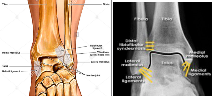
What are your landmarks?

The goal is to avoid the Dorsalis pedal artery, the peroneal nerve and the tendon of the Extensor Hallucis Longus (EHL). It is recommended to use an anterolateral approach where the joint line can be found between the lateral edge of the EDL and the medial edge of the lateral malleolus (Yellow Arrow Image 1). Plantarflex the ankle while the patient is bent at the knee in the supine position to widen the joint space prior to performing the procedure.

IMAGE 1:

Foot.jpg


How do you perform it?

  • 1. Patient should be in a supine position with the ankle in plantar flexion with plantar surface flat on the bed.  

  • 2. Mark you landmarks (see above).

  • 3. Prepare the site (ex. chloraprep)

  • 4. Anesthetize the area with smaller needle(23/25 gauge) creating a wheal and then advance creating the start of a projected path towards the joint capsule.

  • 5. Attach a 5 or 10 cc syringe to a 20 or 22 gauge needle and advance the needle into the joint space pulling negative pressure as you advance. The needle should be directed perpendicular to the tibia. If your syringe starts to fill up, and you need to get more fluid out, change out your syringe using hemostats to hold the needle. Most wrist and ankle effusions will yield only 1-3cc of fluid.

What about Ultrasound Guidance?

YES. This can absolutely be used to assist you in performing the procedure and will allow for visualization of your needle tip during aspiration.


For ultrasound guidance an anteromedial approach is generally used.

Landmarks- Place probe in between the TA tendon and EHL tendon, then rotate longitudinally with the probe marker facing the patient’s head  (Blue Arrow IMAGE 1). You will actually be inserting your needle medial to the TA tendon (Red Arrow IMAGE 1).

Image 2: 

more-tips-and-tricks-6.jpg
more-tips-and-tricks-7.jpg

Image 3:

AnkleTap.png

Ankle fractures

Ankle fractures:


Start with good H&P:

History:

Mechanism, height of the fall, direction of the foot inversion

Consider age, steroid use, hx of neoplasm, prior surgeries, hardware


Physical:

Start from the knee down, neurovascular intact, ROM, strength, severe tenderness, instability, rash/ulcers

Ottawa Ankle rules 


Classification of the injury: stable/unstable?

Many classifications are available but for ED we can use Closed Ring System: 

image8.jpg

Think of an ankle as a ring of bone and ligaments surrounding the talus consisting of the tibia, the medial malleolus and medial deltoid ligaments, the fibula and lateral ligaments and calcaneus.


A single disruption in the ring - stability most likely preserved

Two disruptions - think instability and will likely cause the joint to shift.

Exceptions: Lateral malleolus fracture even with no medial injury may become unstable.

Isolated syndesmosis injury


Approach to ankle injuries x-ray interpretation

Here is an EM focused summary

Look at the cortical disruption of each bone

Look at the soft tissue swelling

Look at the spaces between the bones

Look within the bones

Ask for a mortise view (no, it’s not a GOT character) in addition to the standard AP and lateral views

Look at the tib/fib, knee and base of the 5th metatarsal

Key areas:

Talar shift: look to make sure there is congruence between the clear space on either side of the talus; go further - measure the medial clear space and the lateral clear space. If they are incongruent or the medial clear space is >4mm the ankle is likely unstable.

Talar tilt: The lines in red below should be parallel. Talar tilt indicates an unstable ankle 


Just a few commonly missed fractures at the ED:


High ankle sprain: The isolated syndesmosis injury - isolated distal tibiofibular syndesmosis injury, with ligamentous disruption can result in unstable ankle injury.

Look at the tibio-fibular clear-space: Measure the gap between the tibia and fibula 1cm proximal to the tibial plafond on both the AP view and mortise view. They should be <6mm. If  >6mm, suspect a syndesmosis injury.

Tillaux fracture - fracture is an intra-articular Salter-Harris class III fracture of the distal tibia with avulsion of the anterolateral tibial epiphysis.

Remember that in children, the ligaments tend to be stronger than the growth plate. Tillaux fractures can be considered “the syndesmosis injury of children


Snowboarder’s fracture - A snowboarder’s fracture is a lateral process of the talus fracture that is commonly misdiagnosed as a simple ankle sprain. 


Lateral process of the talus fracture also known as a snowboarder’s fracture


Bottom line: 

Reassess including the if the pt is still neurovascular intact

If pt can’t ambulate get further workup

If in doubt call radiology

Persistent pain but pt wants to go hoe, splint with ortho follow up


References: CoreEM, EMDoc, Uptodate, Radiopedia


5th metatarsal fracture

5MT.jpg

Normal Apophysis in children runs parallel to the bone.  


Dancer’s or Avulsion (pseudo-Jones) Fracture @ cuboid articulation - hard sole shoe for 4-6 weeks and weight bearing as tolerated (WBAT) with orthopedics follow up in a week

Jones Fracture @ intermetatarsal articulation - high risk of non union, pt will need a splint and non weigth bearing activity (NWB) for 6-8 weeks with orthopedics follow up


Metatarsal shaft fracture - high risk of non union, will need a splint, NWB for 10-20 weeks, with orthopedics follow up

Below is a 5 minute video by amazing Dr. Anna Pickens (former Maimo attending) for visual review of the fractures:


http://www.emdocs.net/em-in-5-5th-metatarsal-fractures/

https://youtu.be/4k1dvPdpW4E