VOTW: Do you know the muffin man?

 ·   · 

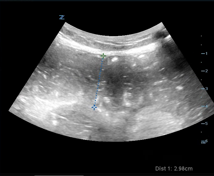
Case: 57 yoF with PMHx of osteoarthritis presented to the ED with right knee pain. A bedside US was performed that showed bulging of the right medial meniscus with surrounding edema Image 1, videos 1&2), consistent with a tear in the medial meniscus.

Meniscal Tear

How to assess the meniscus:

  1. Have the patient flex their knee slightly to 20-30 degrees (you can prop their knee up with a rolled towel) 

  2. Use the linear probe and place it longitudinally along the medial aspect of the knee 

  3. Identify the medial collateral ligament (MCL), which will appear as a hyperechoic and fibrillar structure, extending from the medial femoral condyle to the proximal tibia

  4. The meniscus will appear as a triangular structure that sits between the femur and tibia, under the MCL

5. A meniscal tear is identified with a well-defined anechoic or hypoechoic area surrounding the meniscus. It can cause extrusion of the meniscus as a result of surrounding edema, described as a "muffin top". There may also be increased vascularity in the surrounding area when color flow doppler is applied

Note: The same process can be repeated on the lateral aspect of the knee to assess the lateral meniscus (image 3).

Case conclusion: While the patient had pain with flexion, she was able to ambulate independently. She was discharged with orthopedic follow-up and conservative measures.

Happy scanning! 

Ariella Cohen


References: 

https://theultrasoundsite.co.uk/ultrasound-case-studies/

https://ultrasoundpaedia.com/knee-normal/

https://www.nysora.com/ultrasound-of-the-musculoskeletal-system/chapter14-knee-preview/


Poo-and-fro

 ·   · 

94 yoF with PMHx of gastric cancer and recent SBO (managed non-operatively) presented to the ED with worsening abdominal pain, constipation, and obstipation.

An ultrasound was performed that showed multiple signs consistent with an SBO:


  • Image 1: Dilated loops of bowel > 2.5 cm.

  • Video 1: To-and-for movement of fluid in the bowel. Normally, feculent material should only move in the direction of peristalsis. However, if there is a distal obstruction, you will see feces move back and forth as it attempts to move past it.

  • Video 1: Keyboard sign - when the plicae circulares, finger-like projections of the jejunal inner wall, become more prominent during an obstruction.

SBO

Other sonographic signs of SBO include:

  • A thickened bowel wall > 3 mm.

  • Free fluid between the loops of bowel. 

  • Decreased/absent peristalsis. (Note: Free fluid between bowel loops and lack of peristalsis may indicate bowel ischemia and a worse prognosis.) 

Case conclusion: CT scan was done that showed a distal small bowel obstruction. The patient was admitted to SICU for serial abdominal exams and non-operative management of her SBO.

Happy scanning!

- Ariella Cohen, M.D.


References

  1. https://www.emdocs.net/us-probe-ultrasound-for-small-bowel-obstruction/#:~:text=%E2%97%8B%20Jejunum%20will%20have%20%E2%80%9Cvalvulae,known%20as%20the%20keyboard%20sign).&text=%E2%97%8B%20Ileum%20will%20not%20have%20haustra%20or%20valvulae%20conniventes.&text=Look%20for%20compressibility.

  2. https://coreultrasound.com/small-bowel-obstruction/

  3. https://www.emhum.com/?p=472

 · 
Share

VOTW: Subchorionic Hemorrhage

 ·   · 

Case: A 26 yoF who was 5 weeks pregnant presented to the ED after being pushed out of a parked car by her partner. She complained of wrist pain but requested an US to check on her pregnancy. She denied vaginal bleeding or pelvic pain. She had an IUP that was confirmed via US before the assault.

A transvaginal US was performed (video 1) that showed a subchorionic hemorrhage (SCH) > 50% of the gestational sac. Notebe careful not to confuse a large SCH with another gestational sac (image 1). 

Image 1

A SCH typically occurs within the first 20 weeks of gestation and is when blood accumulates between the uterine wall and the chorionic membrane (image 2). While many are found incidentally, some patient's may present with vaginal bleeding.

(What is the chorion? It is a membrane that surrounds the developing fetus along with the amnion. It eventually forms the fetal placenta and provides nourishment and protection for the developing embryo.)

On ultrasound, a SCH will typically appear as a crescentic collection with an elevation of the chorion. The echotexture can vary from hyperechoic (acute) to hypoechoic (chronic) based on the duration of the SCH. A SCH is considered large if it is > 50% of the size of the gestational sac. While many resolve during pregnancy, a large SCH can increase the risk of placental abruption, preterm labor, and miscarriage. These patients therefore require close OB follow-up for serial ultrasounds.

Case conclusion: the patient’s workup was negative, she was educated about the found SCH, and given OB follow-up later that week. 

Happy scanning!

Ariella Cohen, M.D.


References:

https://radiopaedia.org/articles/subchorionic-haemorrhage-2?lang=us

https://my.clevelandclinic.org/health/symptoms/23511-subchorionic-hematoma