VOTW: Throw what you know

This VOTW is brought to you by Drs. Chiu, Butt, Burns, Wong, and Sanghvi on a scan shift. 

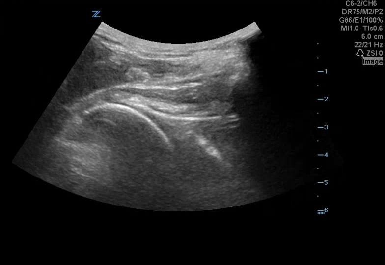
A 42 yoM presented to the ED with a left shoulder dislocation. The ultrasound team looked at his dislocated shoulder (Image 1) and gave an Intraarticular lidocaine injection. The providers reduced his shoulder and then looked for confirmation using ultrasound (Image 2).


How can I do this?

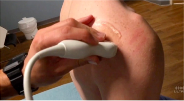
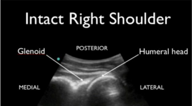
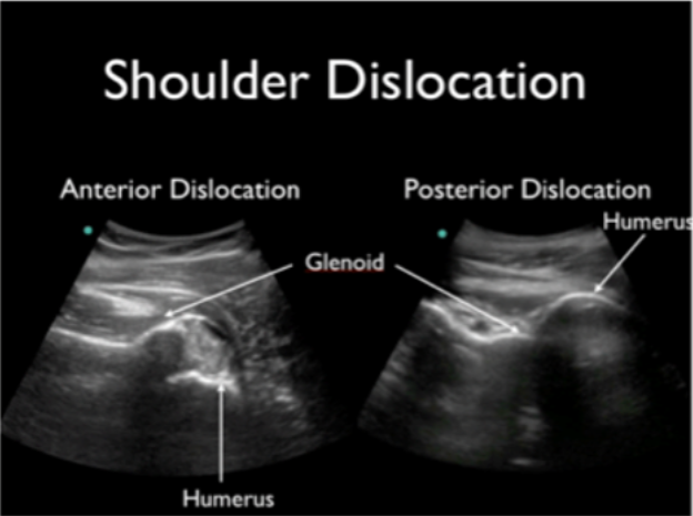
Take your linear or curvilinear probe and place it in transverse orientation on the patient’s back next to the humerus (image 3). The glenoid should articulate directly with the humeral head (image 4). In an anterior shoulder dislocation, the humeral head will be deeper on your screen because it is further from the probe. In a posterior shoulder dislocation, the humeral head will appear more superficial because it is closer to your probe (image 5).

Why use ultrasound?

Ultrasound allows you to check in real-time whether or not the reduction was successful, rather than waiting for x-ray confirmation. I find this particularly useful for my workflow in cases where I am not 100% certain that the shoulder is back in.

References:

  • Martinoli, C. (2010). Musculoskeletal ultrasound: technical guidelines. Insights into imaging1(3), 99.

  • Jacobson, J. A. (2011). Shoulder US: anatomy, technique, and scanning pitfalls. Radiology260(1), 6-16.

  • 5 Minute Sono

  • The Pocus Atlas

Happy scanning!

Ariella Cohen, M.D.


Intubating Asthmatic Patients

Asthma is Greek for panting, which is a fitting translation for a patient that presents with a severe asthma exacerbation. We try to avoid intubating these patients because they are prone to compilations such as pneumothorax, mucus plugging, and increased morbidity and mortality. 

However, there are specific situations when you may consider intubating an asthmatic patient. One reason is that your patient may not be improving despite maximal medical therapy, such as BIPAP, albuterol, ipratropium, magnesium, epinephrine/terbutaline, ketamine, etc. Another reason is that your patient may now be altered, and have worsening work of breathing, and vital sign abnormalities. Remember that a “silent chest” is a poor prognostic indicator; you may not hear wheezing because they are not moving any air. 

If you choose to intubate, there are tricks to maximize your success and optimize your management of your patient on the vent. 

  • Use a large ETT (8-9) because it reduces airflow resistance and can facilitate procedures later (such as bronchoscopy). 

  • Ketamine is a useful induction agent because of its bronchodilatory effects. It may also be useful if you choose delayed sequence intubation. 

  • High airway pressures can cause hypotension after intubation, so consider giving volume if there is a current or prior history of hypotension. 

  • If hemodynamics are compromised consider giving an epinephrine drip. It is considered a systemic bronchodilator that can provide hemodynamic support as well as bronchodilation. 

  • Keep a low respiratory rate when bagging or on the vent (6-8 breaths/min). Giving them time to exhale will decrease the chances of air trapping and pneumothorax. Another way to do this is to increase the I:E time (1:4 or 1:5). 

  • If the vent is alarming, troubleshoot (DOPES mnemonic) but be suspicious for mucus plugs, pneumothorax, or breath stacking. If they are breath stacking, disconnect them from the vent and push on their chest to help them fully exhale.  

A quick note about auto-PEEP and breath stacking: Auto-PEEP refers to trapping gas in the lungs during respiration. This occurs when one breath can’t be fully exhaled before the next inhalation. This trapped gas causes additional positive pressure, known as “auto-PEEP” in the chest which is typically higher than the PEEP set on the ventilator. This process predisposes patients to develop a pneumothorox. 

Thanks for reading!

Ariella


Unstable Pelvic Ring Fractures

The pelvic ring consists of the sacrum and two innominate bones, which are made up of the pubis, ilium, and ischium. These bones are held together by strong ligaments to give the pelvis stability.

A pelvic ring fracture is a severe fracture with 2 breaks in the circular ring, leading to an unstable pelvis and a potentially unstable patient. Fractures that disrupt the pelvic ring predispose patients to bleeding given the large network of arterial and venous anastomoses. Patients who have an isolated pelvic fracture and are hypotensive carry a mortality of 15-40%. Most vascular injuries in the pelvis are venous (90%). While rare, arterial bleeds (10%) should be suspected when a pelvic binder is placed but the patient remains hemodynamically unstable. The retroperitoneal space can accumulate 4 liters of blood before venous tamponade occurs. Pelvic binders are useful in that they can help tamponade bleeding veins, decrease total pelvic volume, and prevent the shifting of bony fragments.

Other unstable pelvic fractures include lateral compression fractures, "open book" pelvic fractures, and vertical shear fractures. Lateral compression fractures occur when a lateral force vector (t-bone in an MVC) causes an anterior ring disruption and sacral fracture.

“Open book” fractures occur as a result of anteroposterior compression injury to the pelvis, commonly caused by high-speed trauma or elderly falls. There is a disruption to the pubic symphysis and the pelvis opens like a book. Diastasis of > 1 cm (blue arrow) can indicate instability. Disruption of the pubic symphysis, one of the strongest ligamentous structures in the human body, requires a lot of force and should be a red flag to look for other injuries to the head, spine, chest, or abdomen.

Vertical shear pelvic fractures are seen when one-half of the pelvis shifts upward as a result of a fracture of ipsilateral anterior and posterior pelvic ring fractures. They typically occur as a result of high-energy force applied in the axial direction (aka from the gas pedal to the femur and up to the pelvis). Patients may have an unstable pelvis and leg length discrepancy.

For all unstable fractures, you should appropriately resuscitate and stabilize the patient. Give blood as needed but avoid transfusing through lower limb access because it may drain into the retroperitoneal space. If there is a pelvic ring fracture, consider binding the pelvis. Your binder should lay over the greater trochanters and have enough force to close the pelvic ring (video:https://www.youtube.com/watch?v=tWLBZKeWEkg).