Diplopia

Diplopia

Case

48F pmh htn, DM presenting with difficulty focusing on distant objects for past few days. Feels like she sees double, all other review of systems normal.  On physical exam, pt has a 6th nerve palsy, but has limited R sided rapid alternating movements. CT/MRI/MAR performed shoulder nonhemorrhagic pontine infarct.

Evaluation

1. Is it monocular diplopia or binocular diplopia? -> have patient cover each eye and see if diplopia persists

If diplopia resolves when either eye is covered -> binocular diplopia

If covering one eye resolves the diplopia -> monocular diplopia in the covered eye

Monocular diplopia – refractive error of eye, retinal disease, less common CNS pathology though possible

              Usually does not require neuroimaging, can send for optho referral

Binocular diplopia – neuromuscular dysfunction causing misalignment of visual fields, CN palsy, oscular muscle dysfunction, brainstem pathlogy

              Usually needs further ED workup for neurologic problem

2. Is diplopia isolated or associated with neurologic signs? Full neuro assessment/exam.

Is there ataxia, vertigo, neuro deficits, dysphagia/dysarthria -> needs rapid neuroimaging and stroke consult to r/o stroke, ideally should have MRI to assess posterior circulation

3. Try to localize lesion casing diplopia especially 3rd and 6th nerve palsies by having patient look horizontally and vertically

3 6 palys.JPG

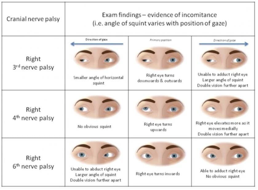
Isolated nerve palsies are the most common causes of diplopia usually caused by microvascular insult of the extraocular muscles

Isolated 3rd nerve palsy always needs NCHCT and CTA as they are often caused by ischemia or compressive aneurysm

Isolated 4th nerve palsies without other concerning findings can follow with neurology

Isolated 6th nerve palsies usually does not need imaging unless concern for mass/increased ICP, pediatric 6th nerve palsy needs imaging as malignancy is more prevalent than microvascular disease

If there is suspicion of trauma, pt need neuroimaging regardless of whether it’s monocular or not, orbital floor fracture can entrap extraocular muscles or cause a retrobulbar hematoma causing diplopia

Other Diseases to watch for

Cavernous sinus thrombosis – occlusion of cavernous sinus housing CN 3,4,6, often have eye of affected eye, headache, risk factors for thromboembolic disease -> need CT venogram and MRV

Thyroid disease – cause enlargement and fibrosis of extraocular musculature, fatigue, wt loss, TSH/T3/T4

Giant Cell Arteritis - > 50 yo, unilateral HA, ESR/CRP

Myasthenia Gravis – proximal wkns, respiratory distress, fatigable rather than persistent

4. Patients should be dced with neuro/optho f/u as there are many complex motility disorders that need non-emergent MRI or detailed optho exams


Needle Insufl

POTD: Jet Insufflation

Case:

3M with peanut allergy with rash and difficulty breathing. The child was just watching TV when he started itching then developed rash over his entire body and is struggling to breath.  Epi pen given but the patient isn’t improving.

PE: severe distress, angioedema in eyes, lips, tongue, stridor and wheezing heard, tachypneic and accessory muscle use, severe urticaria skin

IOs are placed, started on NRB, epi given, Benadryl given, steroids, albuterol administerered

Pt mental status rapidly decreasing becoming cyanotic, BVM attempted but poor air entry, quick attempt at DL shows significant airway edema with no view of the cords…what next

Great 2 min video going over how to perform Needle Cric

https://www.youtube.com/watch?v=vOG08zTZ2YE

Background/Indications

Percutaneous transtracheal jet ventilation is indicated for pediatric patients who cannot be ventilated with BVM or intubated whether due to severe maxillofacial trauma, upper airway bleeding, mass, severe swelling/edema, burns, etc. 

Preferred surgical airway for children under 12yo due to small tracheal diameter, prominent hyoid bone which prevents open cricothyrotomy

Not a definitive airway, only a temporizing measure ideally < 30 min until definitive airway is obtained (bronchoscopy, surgical tracheostomy)

Contraindications – significant damage to cricoid cartilage or larynx, if endotracheal intubation is possible

Relative contraindication – complete upper-airway obstruction (surgical cricothyroidotomy preferred

Equipment

O2 source at 10-15L/min

BVM

Large-bore, over the needle IV cath (18 ga+)

Sterile gloves and prep

Depending on what’s available:

              3.0 ETT hub (hub is the end of the ETT that connects to the BVM/vent that can be removed)

              1 cc syringe + 3.5 ETT Hu

              3 cc syringe + 7.5 ETT Hub

              Jet Insufflation Kit

1. Ideally, you have time to sterilize the field while the patient is supine

2. Identify the cricothyroid membrane, there are blood vessels sometimes overlying the upper third of the cricothyroid membrane, puncture the lower third if possible

3. Using the largest bore IV, remove the needle portion with the cath

4. twist on a 10 cc syringe (5cc ok) and fill half way with saline

5. stand at the head of the bed and aim the needle caudad at 30-40 degree angle, aspirate as you advance slowly to prevent puncture of the posterior tracheal wall, when you visualize air bubble drawback stop

6. once in the trachea, slide the catheter into the trachea, ideally verify placement in trachea again by aspirating with your 1 cc or 3 cc syringe

7. place either your 3.0 ETT hub directly on the end of the catheter or the 1 cc or 3cc syringe hub combo

8. connect the ETT hub to a BVM

9. suture your catheter in place and plan for definitive airway measures

In our Peds ER, we have a Jet Insufflation kit in the top shelf of the R cabinet in Peds resus.  There are three things in the bag: needle, syringe, and tubing. Prep the patient as you normally would. 

1. attach saline filled syringe to needle, insert needle same as a standard needle cric while checking for bubble

2. once in trachea, remove syringe and advance the catheter

3. connect the tubing to the end of the catheter, the soft plastic end should be connected to the wall oxygen 10-15L/min

3. Note in the second image, there are holes in the side of the tubing connected to the needle/catheter, these need to be covered for air to go to the patient.  You will be controlling the patient’s breathing.

You will be giving breathes at a rate depending on the patient’s age and pathology; the I:E ratio should be usually 1:4 to 1:5

I to E ratio is inspiratory time to expiratory time ratio (same as a vent)

Ex: you want a RR of 20 so each complete breathe is 3s, I:E of 1:5 would be inspiration time of 0.6s with expiration of 2.4s.  Of course, in real life, manually trying to time 0.6s inspiration etc is impossible but just try to keep in mind that the time you are giving air should be shorter than the relaxation/exhalation phase. If you ended up giving 1:1 ratio, you might end with a hypercarbic, breath stacked, barotrauma patient.

mc1.jpg

Electric Injuries

Electrical Injuries

Starting off, an electrical injury patient is a trauma patient first, make sure to follow primary and secondary survey, EFAST, etc.

May require intubation if obtunded

r/o TBI, spinal cord, blunt thoracic, etc.

Risk Stratification

Low voltage (<600V) – household or office exposures lower risk injuries

High voltage (>600V) – industrial settings, subway rails, power lines are high risk injuries

Alternating Current (AC) – causes prolonged contraction and release of muscle which prevents full release from electrical source which will cause longer contact duration and more tissue damage compared to Direct Current (DC)

Lightning strikes (up to 1 billion V) are DC with brief contact

80% have long term morbidity, 10-30% mortality

Asystole from depolarization of myocardium but sponteous ROSC often achieved

Respiratory arrest from medullary paralysis

For lightning strike mass casualty incident, resuscitate those who appear dead first

Wound Care – saline moistened gauze (ideally sterile) and antibiotic ointment (silver sulfadiazine cream, bacitracin, mupirocin) unless transferring to burn center which mostly prefer just sterile moistened gauze

Cardiac Complications

Bundle branch blocks, AV blocks, QT prolongation, ST changes, Afib – most resolve spontaneously, Vfib (more with AC, asystole more with DC)

STEMI can arise rarely from electrical shocks, can consider trop

Compartment Syndrome – need to monitor, especially burn sites

Rhabdomyolysis/AKI

CK correlates to extent of muscle injury

Can have tea colored urine

Can have hyperK from AKI/Rhabdo but usually resolves with fluids

Treat as normal rhabdo with IVF and consider urine alkalinization with bicarb gtt

Electrical cord bite injury

Children can bite on electrical cords causing damage to oral pharynx, delayed massive bleeding from labial artery can arise – can grip lips with fingers/gauze to decrease bleeding

Cardiac Monitoring

Low voltage exposure w/o chest pain/syncope, no need for ECG monitoring

High voltage exposure – cardiac monitor 6-8hr

Delayed complications – altered mental status, memory loss, limb ischemia from vasospasm, parasthesias, cataract formation, delayed anerusym formation, delayed thrombosis

Disposition

Asymptomatic low voltage can be discharged

High voltage obs for 12 hrs with 6 hrs cardiac monitoring

Admit for dysrhythmia, AMS

Refer to burn center if significant burn injuries