Stop Giving Amps of Bicarb!

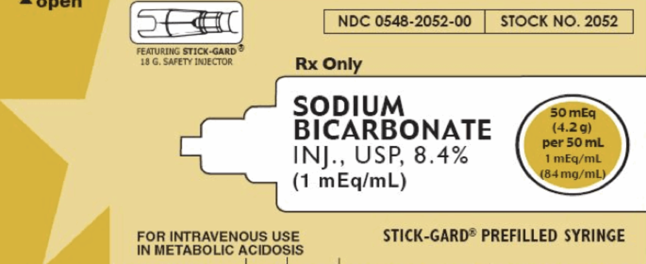
Chapter 1: What dafuq is in an amp of bicarb?

Take a look!

  • 50mL

  • 8.4% NaHCO3 -> 50mEq

  • The osmolarity of this solution is 2,000mOsm/L - twice that of 3% saline. < (click for emcrit)

Screen Shot 2020-11-05 at 7.56.02 PM.png

Chapter 2: Sodium bicarbonate doesn't just magically raise pH...

Remember this thing?

CO2 + H20 <=> H2CO3 <=> HCO3 + H

It's complicated. Bicarb binds to acid. Then it turns to CO2 and water, so you can breathe it out.

Basically if you're giving bicarb, you can only raise your pH as long as you can breathe off your CO2, increasing your RATE or VOLUME.

**This is particularly a problem in patients who are not in control of their breathing (vented), aren't breathing (arrest), or who have maximized the efficiency of their breathing (Kussmal breathing in DKA).**

That's right - you need to increase your minute ventilation to have a change in pH.

Here's Weingart's take.

Chapter 3: Sodium bicarb amps can cause harm!

FIRST:

One amp of bicarb is like giving 100cc of 3% hypertonic saline!! But as Josh Farkas points out, we typically have no hesitation giving "a couple of amps of bicarb."
This is a huge osmotic load which can lead to huge fluid shifts - prepare for that amp to increase intravascular fluid by 1/4 liter with every push. (Is this what you want to give to your renal failure pt? Your heart failure pt?)

SECOND:

You are worsening acidosis.
What? Huh? But I thought...
No. Stop. Shush. You're worsening acidosis.

Remember, you're increasing CO2 - whether you can breathe it off or not, this CO2 rises in but blood BUT ALSO rises in the tissues and may worsen acidosis in these tissues. < (click for litfl.com article)

THIRD:

Be ready to cause hypernatremia - expect a rise of 1mEq Na per amp of bicarb.

FOURTH:

Extravasation can cause tissue necrosis.

FIFTH:

CSF acidosis, hypocalcemia. Increased lactate. (Some may argue that's not a bad thing.)

If you do manage to fix the acidosis, you can overshoot and create an alkalosis and even screw up the oxygen dissociation curve (in a bad way).

Chapter 4: It just doesn't f&$%ing work
Cardiac arrest: it doesn't do anything. No increased survival. and AHA says it should not be given routinely.

Lactic acidosis: There's a whole section on UpToDate - there's minimal research for pH < 7.1 so you can consider it at that point... but otherwise, nah.

DKA: Take it from a nephrologist: In ketoacidosis, it is almost never necessary to give bicarbonate even though the patient is bicarbonate deficient unless renal function is permanently impaired. Therapy with fluids and electrolytes restores extracellular volume and renal blood flow, thus enhancing the renal excretion of acid and regenerating bicarbonate.

Hyperkalemia: Amps of bicarb, even in hyperK emergencies, have not been shown to lower potassium. Click that UpToDate link or listen to Scott Weingart talk about it on EMRAP.
Patients with hyperK should be started on isotonic bicarbonate drips for 4-6hours, a treatment that works better in acidotic patients.

CHAPTER 5: Soooo who gets bicarb?
AMPS:

  • Bicarb ampules in sodium channel blockade (like TCAs) are, as Dr. Bogoch said yesterday, the cornerstone of therapy

  • Bicarb ampules may be appropriate to alkalinize urine in certain toxicities

  • Seizing hyponatremic patients

DRIPS:

  • Appropriate in hyperK patients who can handle fluid

  • Appropriate in patients with AKI and pH < 7.2 (BICAR-ICU Trial)

  • May be appropriate for pH < 7.0 or 7.1, depending on who you talk to...

**If the pH is < 7.1 and you wanna give an amp of bicarb, there isn't enough data to say you're wrong. If it's a last-ditch effort, you might as well.

https://www.uptodate.com/contents/bicarbonate-therapy-in-lactic-acidosis?search=sodium%20bicarbonate&source=search_result&selectedTitle=3~148&usage_type=default&display_rank=2

Other references embedded in text.


Presidential Pathology

In honor of election season, let’s review some pearls and boards material surrounding our nation’s presidents!

Woodrow Wilson – Influenza, Stroke

1918: There is some suspicion that President Wilson caught the famous virus from the 1918 flu pandemic. He later suffered a TIA and a massive stroke (L hemiplegia) – his staff hid the severity of his stroke while his wife supervised his duties.

Influenza:

·      Antigenic drift = small mutations that create different seasonal flus

·      Antigenic shift = switches species

·      Tamiflu = all hospitalized & high risk patients ASAP, low risk patients within 48hrs

o   Tamiflu debate, click here

Stroke:

·      Highest risk of stroke after TIA = 48hrs

·      Blood pressure goals:

o   Ischemic stroke, TPA eligible = Keep below 185/110

o   Ischemic stroke, no TPA = 220/120

·      TPA to be given within 4.5 hours


FDR – Polio

1933-1945: Photographers avoided taking pictures of FDR while he was in his wheelchair as it was viewed as a sign of weakness. Photos of him were deliberately taken only while the president was in a car or behind a desk.

Polio

·      The WHO anticipated eradicating polio from the planet by 2023. However, President Trump’s withdrawal from the organization had led to a severe decrease in funding and that may need to be reconsidered. Check out this clip from Sunday’s Last Week Tonight With John Oliver to learn more.


Eisenhower – MI, Crohn’s Disease

1955: He stayed in Fitzsimons Army Hospital in Colorado for 7 weeks after his heart attack, but I couldn’t find how they treated it. Just a few months later, six months prior to his next election, he was diagnosed with Crohn’s Disease and required surgery. He went on to win the election.

MI: Lysis vs Cath

·      Lysis if PCI cannot be performed in the “appropriate timeframes” below

·      PCI timeframes:

o   AMI within 2hrs = PCI in 60 minutes

o   AMI within 2-3hrs = PCI in 60-120 minutes

o   AMI within 3-12hrs = PCI in 120 minutes

Crohn’s Disease

·      Typically 2nd/3rd decade of life, male, hx of IBD in the family

·      Any part of the digestive tract from mouth to anus

·      Skip lesions

·      Full thickness inflammation (unlike UC = epithelial layer only)


Jed Bartlet – Multiple Sclerosis

2000: Widely acclaimed as the greatest president of our time, the Bartlet Whitehouse was rocked by scandal and outrage when it was revealed that the president and members of his administration had willfully omitted knowledge of the president’s devastating demyelinating disease. Despite the controversy, Americans saw past this lapse of judgement and reelected President Bartlet for a second term.

MS

·      Autoimmune, more in females, connected to psoriasis and thyroid disease

·      Internuclear ophthalmoplegia = difficulty adducting eye = pathognomonic

·      LP = Oligoclonal bands & IgG in the CSF

·      MRI = optic nerve lesions, juxtacortical lesions, and Dawson Fingers

·      Steroids for flares (inpt or outpt)


George W. Bush – Colonoscopy

2002, 2007: Colonoscopies aren’t that interesting but Bush did, indeed, hand over the power of the presidency to Dick Cheney on two occasions, each lasting just over 2hrs while he had routine colonoscopies.

Colonoscopy

·      Q10yrs, starting at age 50 (unless family hx, familial adenomatous polyposis, etc.)

·      Complications:

o   Pyogenic liver abscess

o   Infection

o   Bleeding (post-polypectomy, 1 week after procedure)

o   Perforation

o   Post-polypectomy syndrome: peritonitis without perforation after a transmural burn in the colon


Kennedy – Addison’s Disease

1961-1963: It looks like JFK suffered from quite a number of medical problems: chronic back pain, colitis, UTI, abscess, possibly malaria, and apparently was on a brief course of antipsychotics after a change in mood when he started some antihistamines. The most famous of these maladies was his Addison’s Disease, for which he was on daily steroids.


Interestingly, Kennedy was wearing his back brace on the day he was assassinated, which kept his posture fully upright in the limousine prior to getting shot.

Addison’s Disease:

·      Chronic adrenal insufficiency, autoimmune – patient’s on chronic steroids

·      Hyperpigmentation

·      Must be distinguished from acute adrenal insufficiency:

o   Look for hyponatremia and hyperkalemia (low aldosterone)

o   Hypoglycemia

o   Refractory hypotension

o   Hydrocortisone 100mg IV


REFERENCES:

https://www.cnn.com/2020/10/07/health/us-presidents-health-problems-wellness/index.html

https://www.healthline.com/health/diseases-of-presidents

https://www.ahajournals.org/doi/10.1161/STR.0000000000000211

https://text-message.blogs.archives.gov/2016/09/22/heart-attack-strikes-ike-president-eisenhowers-1955-medical-emergency-in-colorado/

http://www.emdocs.net/multiple-sclerosis-ed-pearls-pitfalls/

https://www.businessinsider.com/25th-amendment-colon-trump-reagan-bush-unfit-president-2017-10

http://www.emdocs.net/post-colonoscopy-complications/

peerix.acep.org


T Wave Inversions

EKG#1

2yoF with fever. There was a miscommunication and someone accidentally got this EKG – even no one asked for it. Many emails have been sent as a result, swaths of staff fired and ridiculed… but even still, you’re stuck with this EKG.

1. Is this 2yo having an MI? Spontaneous coronary artery dissection? Coronary aneurysm from Kawasaki?

1. Is this 2yo having an MI? Spontaneous coronary artery dissection? Coronary aneurysm from Kawasaki?

EKG#2

48yoM with exertional chest pain. He’s wearing a fedora and sunglasses indoors. Is this clinically relevant? You decide.

1. Iunno, what do you think? Good? Bad?

1. Iunno, what do you think? Good? Bad?

EKG#3

88yoF found unresponsive in the bathroom. No cardiac history.

1. Which came first, the EKG or the fall?

1. Which came first, the EKG or the fall?

EKG#4

33yoM, exertional SOB

1. Do you agree with the computer interpretation?2. What test will your order next?

1. Do you agree with the computer interpretation?

2. What test will your order next?

PRECORDIAL T-WAVE INVERSIONS (TWI) EDITION

What’s the differential?

Wow, look at this snazzy graphic you can reference!

TWI5.png

Before we start…

…let’s review normal T waves.

Screen Shot 2019-11-26 at 4.19.28 PM.png

T waves should be…

1.   Upright in all leads except aVR and V1 (sometimes V2)

2.   Asymmetric, with a gradual upslope and a steep return to baseline

3.   Smaller than the QRS


Lots of things can invert your T waves

·      We will focus today on the differential on the first page

·      In addition to these dangerous pathologies, consider…

o  Ventricular strain

o  HCM

Lead placement:  https://litfl.com/ecg-limb-lead-reversal-ecg-library/

ANSWERS 

EKG#1

2yoF with fever – Juvenile T Waves

·      Remember, TWI is a normal finding in children.

·      Note that these TWI’s are asymmetric, as opposed to the next pathologic example.

o   They have a gradual upslope, steep downslope

·      They may become upright as early as age 8, or they may “persist” into adulthood.

·      Persistent Juvenile T Waves (PJTW) typically present African American women <30yo

but…

·      As Dr. Richard Wang said, PJTW is a diagnosis of exclusion. It’s reasonable to interpret this TWI pattern as normal in a child, but consider this pathologic on the adult side until proven otherwise.

From Richard:

·      Remember right axis deviation is normal in pediatrics – remember that the R side of the heart does most of the work in-utero, so it’s normal to expect it to be [relatively] bulkier

·      This EKG shows “early transition,” meaning R > S in V1/2, suggestive of increased work in the R side of the heart


Additional Reading: https://pedemmorsels.com/pediatric-ecg/

EKG#2

48yoM with exertional chest pain – Wellens, Type B

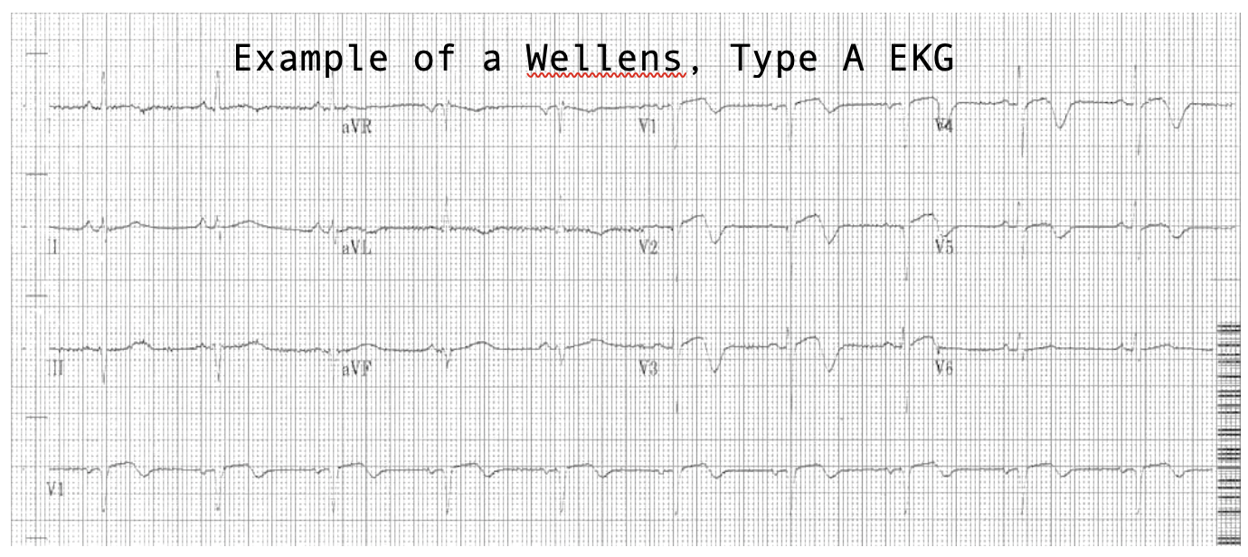
·      Deep, symmetric TWI in the precordial leads = Wellens, Type B

·      There are two types of Wellens patterns, A and B.

·      Both indicate critical stenosis of the LAD and these patients should be treated as impending STEMI’s, though their ST segments may appear normal.


Here is a fantastic explanation of Wellen’s physiology from Life in the Fast Lane:

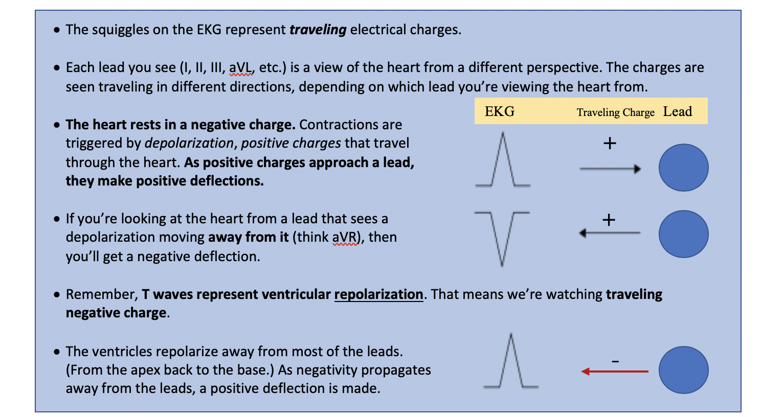
·       A sudden occlusion of the LAD, causing a transient anterior STEMI. The patient has chest pain & diaphoresis. This stage may not be successfully captured on an ECG recording.

·       Re-perfusion of the LAD. The chest pain resolves. ST elevation improves and T waves become biphasic or inverted. The T wave morphology is identical to patients who reperfuse after a successful PCI.

·       If the artery remains open, the T waves evolve over time from biphasic to deeply inverted.

·       The coronary perfusion is unstable, however, and the LAD can re-occlude at any time. If this happens, the first sign on the ECG is an apparent normalisation of the T waves — so-called “pseudo-normalisation”. The T waves switch from biphasic/inverted to upright and prominent. This is a sign of hyperacute STEMI and is usually accompanied by recurrence of chest pain, although the ECG changes can precede the symptoms.

·       If the artery remains occluded, the patient now develops an evolving anterior STEMI.

·       Alternatively, a “stuttering” pattern may develop, with intermittent reperfusion and re-occlusion. This would manifest as alternating ECGs demonstrating Wellens and pseudonormalisation/STEMI patterns.

Screen Shot 2019-11-26 at 4.24.48 PM.png

Additional Reading: https://litfl.com/wellens-syndrome-ecg-library/

EKG#3

88yoF found unresponsive in the bathroom – Cerebral T Waves

·      Deep, SYMMETRIC TWI… are we sensing a pattern here?

·      This is a pretty rare phenomenon that occurs with stroke or increased ICP (think bleed).

·      This particular EKG is from a patient with a subarachnoid hemorrhage.

·      The pathophysiology is currently not known.

·      One study (https://www.ajconline.org/article/S0002-9149(17)31597-7/fulltext) found 2% of stroke patients had inverted T waves, and 18% of those had transient wall-motion abnormalities, suggesting that this finding may actually reflect true cardiac dysfunction.

·      It is common for stroke patients to spill troponin, so add ‘em on! 

Additional Reading: http://www.emdocs.net/ecg-pointers-intracranial-hemorrhage/

EKG#4

33yoM, exertional SOB – Pulmonary Embolism

·      TWI is more prevalent in PE than S1Q3T3, but still not totes specific
https://www.ncbi.nlm.nih.gov/pmc/articles/PMC5306533/

·      Remember that EKG in PE may be:

o   STONE COLD NORMAL

o   Sinus Tach

o   RBBB

o   New Right Axis Deviation 

·      Great EMCRIT article on how to differentiate AMI from PE:
https://emcrit.org/pulmcrit/two-ekg-patterns-of-pulmonary-embolism-which-mimic-mi/


References

https://litfl.com/ecg-changes-in-pulmonary-embolism/

http://ems12lead.com/2014/11/18/anterior-t-wave-inversions-and-pe/#gref

https://litfl.com/paediatric-ecg-interpretation-ecg-library/

https://litfl.com/t-wave-ecg-library/