Don't be Rash about Deadly Rashes

So, let's talk about why you went into Emergency Medicine - to talk about rashes, of course! Although you might not always (or rarely?!?) know the exact etiology of the rash, there are some Can't-Miss life threatening rashes that we must be able to recognize in the ED. And rashes account for 5% of all ED visits, so you should be diligent to identify these dangerous diagnoses. 

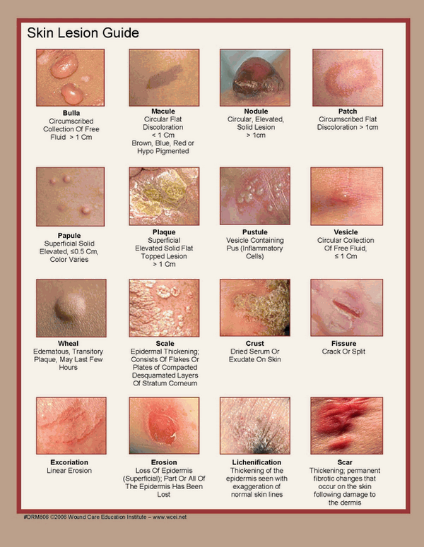
First things first: Describe the rash 

rash pics.png

Pertinent points on managing rashes: 

- STOP IT! Stop the offending agent. Known drugs that induce SJS-TEN include sulfa drugs, penicillin, barbituric acids, aspirins, pyrazolone drugs, and anticonvulsants.

- Rashes can be like a BURN, resulting in abnormal fluid balance, thermoregulation, infection control, and electrolytes. Treat appropriately!

- Treat their PAIN (Make Motov proud!) 

- Sepsis management (Make Dickman proud!). If underlying pathology is infectious, treat early and treat appropriately

- Utilize consultants, the sooner the better! Necrotizing infections - surgery. Toxic rashes - Dermatology. ICU and Burn Centers should be on board!

- Steroids??? WAIT  -  let Dermatology/Burn Center/ICU determine the disease pathology so that steroids will not be given that may cause more harm (SJS/Tens) than good. 

There's so much information, but this is just a rough guideline. Remember to rule out life threatening rashes before sending your patient home. 

You have to determine the TYPE of rash. The algorithm divides the rashes into 6 types: Maculopapular, Petechial/Purpura, Diffuse Erythematous, Non-erythematous, Vesiculo-bullous, Pustular. 

Then branch each type of the 6 rashes into the life threatening etiologies. 

erythematous.png
vesiculobulbo.png

Keep in mind that this list is not comprehensive, but a guideline to help identify life threatening rash. If the patient looks ill, be concerned. 

Once you identify a life threatening rash, be aggressive and manage appropriately (too many points to address here.) Again, just a guide to look for the DANGEROUS RASHES!. Please see the original reference article attached!

REFERENCES: 

https://www.mdedge.com/emed-journal/article/71662/dermatology/emergent-diagnosis-unknown-rash-algorithmic-approach

https://jetem.org/em_derm_tbl/

http://www.emdocs.net/dont-rash-emergency-physicians-approach-undifferentiated-lesion/

 · 

Add Adenosine to the Flush

You have a patient in SVT, failed vagal maneuvers. Time to treat with adenosine. 

You all know this cute little three-way stop cock. Seems simple enough. That is until you need to use it... the stop and go seems somehow far more confusing than it really is.

3w.png


And the one time you MUST know how to use it is to rapidly administer adenosine. You need access in the antecubital or proximal upper extremity.

Why the rush? Adenosine is rapidly metabolized by erythrocytes and vascular endothelial cells - so with its 10 second half-life, we have to administer and flush it quickly so it can reach the heart. 

Surely, there has to be an easier way! Well, folks. There is!

Make sure you have your ECG rhythm strip running, zoll pads on the patient, and explain the patient that this might "feel funny"  (as their heart stops for just a wee bit). 

  • Grab a 20-mL (or 30-mL) syringe.

  • Desired dose of Adenosine (6 mg or 12 mg)

  • Draw up the adenosine AND the normal saline in the same 20-mL syringe.

  • Administer via fast IV push

That's it! 

Adenosine is safe and maintains its effectiveness mixed with normal saline. One study even used OI access for conversion of SVT in an infant. 

Only have central access (hemodialysis port, central line)??? Per 2010 ACLS guidelines drop the dosing: 

  • 1st dose: 3 mg (instead of 6)

  • 2nd/3rd doses: 6 mg (instead of 12)

This lower dosing minimized risks of prolonged bradycardia. ALSO - use this lower dosing if the patient is taking dipyridamole or carbamazepine as these two medications potentiate the effects of adenosine.


REFERENCES:

J Korean Soc Emerg Med. 2003 Aug;14(3):224-227 

https://www.resus.com.au/2015/03/26/a-new-way-to-give-adenosine-in-svt/

https://www.aliem.com/2012/12/trick-of-trade-combine-adenosine-and/
Weberding NT, et al. Adenosine Administration With Stopcock Technique Delivers Lower-Than-Intended Drug Doses. Ann Emerg Med 2018;71(2):220-4.

https://acls-algorithms.com/acls-drugs/acls-and-adenosine/comment-page-2/

 · 

Traumatic Hemothorax

Case: 18 y/o M is wheeled in with a stab wound to the left chest. VS: HR 130, BP 95/45, RR 30, SpO2 92% on 15L NRB. Pt is maintaining airway, no tracheal deviation, diminished BS on the left, strong distal pulses. You place a left-sided 36F chest tube with immediate blood return. 

What are the possible etiologies of traumatic hemothorax?

Laceration/injury to the heart, major vessels, intercostal vessels, mammary arteries, thoracic spine, diaphragm or lung parenchyma. 

How reliable is the FAST exam in diagnosing a hemothorax?

Sensitivity is 92-96% however bear in mind that the presence of subcutaneous air or concomitant PTX may obscure the underlying blood.

How much blood must be present to diagnose a hemothorax on CXR?
For upright CXRs, 150-300mL of blood causes blunting of the costophrenic angle. However, most trauma will have their CXR done in a supine position, which has a low sensitivity 35-60%. It may take 1L of blood distributed throughout a supine hemithorax to develop haziness on a supine film!

What defines a massive hemothorax?
Traditionally:

-Immediate drainage of 1.5L (or 15mL/kg) or 1/3 of blood volume
-Drainage of 200mL/h (or 3mL/kg/h) x 2-4 hours plus persistent need for blood products

Other definitions:

Screen Shot 2018-12-12 at 9.39.07 AM.png

How to manage a massive hemothorax post thoracentesis?

Address hypoxia by keeping patient on oxygen and may attempt to position so that affected lung is down (if permitted by lack of other injuries). Resuscitate with 1:1:1 blood products. These patients benefit from thoracotomy in the OR as soon as possible. 

What are the long-term complications of not adequately draining a hemothorax?

Retained hemothorax consisting of clotted blood can form, which is not easily drainable by a chest tube. A traumatic hemothorax is also a nidus of infection; these patients are at risk of developing empyemas.

Screen Shot 2018-12-12 at 9.41.26 AM.png
 ·