Hemothorax, Pneumothorax

 ·   · 

Today, we are going to discuss a little bit about thoracic ultrasound in the setting of Trauma: From Core EM (Resident homework)  https://coreem.net/core/traumatic-hemothorax/ verbatim:

•Ultrasound (Carmody 2011)

◦High sensitivity and specificity for detecting the presence and quantification of fluid in the pleural space

◦Hemorrhage usually contains echogenic swirling material that moves in response to respiratory movement or the heart beat

◦Best identified using a coronal view of the abdomen (RUQ and LUQ) with a low-frequency probe

◦Twice as accurate as Xray in predicting the volume of a pleural effusion

◦Higher sensitivity for detecting concomitant pneumothorax than CXR

What do we mean by a LOW FREQUENCY PROBE?  This is the C-62 which you’re usually using in the FAST exam already.  Higher amplitude, lower frequency ultrasound waves can penetrate deeper into the body (as opposed to our higher frequency L83 which has a better resolution but cannot penetrate the way the C62 can).

As stated above, ultrasound is better than CXR predicting volume of a pleural effusion and for detecting a pneumothorax.  Let’s look at some pictures!

http://www.erpocketbooks.com/er-ultrasounds/trauma-ultrasounds-from-the-ed/: 

This should be a pretty obvious PTX from radiopedia.org (hint:  look at the right ribs and then see if you can follow the air markings all the way up like you can on the left):  

Great picture of the “Bar Code sign” vs “Waves on a beach” for PTX.  When you have lung sliding (e.g. NO PTX), the lung slides against the pleura and the moving part creates the sandy “beach” in the picture while the stationary pleura creates what looks like “waves”.  Here’s a great link if you want a video and more info!  http://rebelem.com/ultrasound-detection-pneumothorax/ 

Once you’ve established that the patient has a hemo- or pneumothorax that is causing distress, you should place a chest tube.  You perform needle decompression IF the blood pressure is unstable and the patient has a TENSION pneumothorax

Here’s a video on how to put in a chest tube:  https://www.youtube.com/watch?v=U618Jte_1Uk

More Pearls for Needle Thoracostomy with cheesy music:  https://www.youtube.com/watch?v=hiax6H5zUcs

 · 

POTD: OB/Miscarriage

 ·   · 

Abortion Types

Cervix
Fetal Heart Tones Open Closed
Present Inevitable Threatened
Absent Incomplete Missed

-If Threatened Abortion, get quantitative beta-hCG and/or progesterone levels to establish viability and risk of ectopy.

>20 weeks=still birth (intrauterine fetal demise)

First Trimester=usually due to chromosomal abnormalities (~70%); also possible:  infection, maternal anatomic defect, immunologic or endocrine factors.  OFTEN DON’T KNOW.

Second Trimester (up to 20 wks)=multiple things, including infection, uterine/anatomic defect, exposure to fetotoxic agent, trauma, maternal systemic disease.   Type I diabetes and Thyroid antibodies may be culprits!  Usually NOT chromosomal abnormality.

Late second trimester/Periviable deliveries=preterm labor (contractions lead to cervical dilation) or incompetent cervix (painless cervical dilation due to cone biopsy, previous dilation, cervical lac during previous vag deliver, or DES exposure).  Uterus unable to maintain a pregnancy.

Mgmt:  Check vitals, get CBC, quantitative beta-HCG, blood type, antibody screen, coags and ultrasound.

Tx:

If complete (everything came out), just follow for recurrent bleed/signs of infxn

If incomplete, inevitable, or miss, ob may do D&C or administer prostaglandins (misoprostol)

If threatened, pelvic rest w/ nothing per vagina.  At increased risk for preterm labor and PPROM.  Should receive RhoGAM if Rh negative.

If second trimester, D&E or high doses of oxytocin or prostaglandins.  D&E is not inducing labor.  Required to use laminaria to force open cervix which can have several complications (uterine perforation, cervical laceration).   Could also induce labor which takes longer but has fewer complications because no instrumentation is involved.

If trying to save (this is OB, not EM):  cervical cerclage until 36-37 weeks if incompetent cervix OR tocolysis if preterm labor.  If a Transabdominal cerclage/TAC, must do C-section.

Difference between Dilation and Cutterage vs. Dilation and Evaculation is time/procedure.  D&C during first trimester, D&E during second trimester because more difficult/involved.

Recurrent pregnancy loss (3+ losses consecutively), check for:

1.  Antiphospholipid Antibody (APA)- low dose aspirin

2.  Luteal phase defect- give progesterone in future pregnancies

3.  TSH for hypothyroidism

4.  A1C for diabetes

5.  Hypercoagulability;  Factor V Leiden deficiency, Russel Viper Venom, Antithrombin III/prothrombin mutation, Protein S and Protein C- tx w/ Enoxaparin or unfractionated heparin, subQ

6.  SLE (lupus anticoagulant, ANA, dsDNA, anticardiolipin Ab which is also see in Syphillis, Behcet's)

Ob-gyn may do a karyotype, hysterosalpingogram or possibly a hysteroscopic or laparotic exploration.  Still send cultures for infection (vaginal, cervix, endometrium)

 · 

Pacemaker Malfunction

 ·   · 

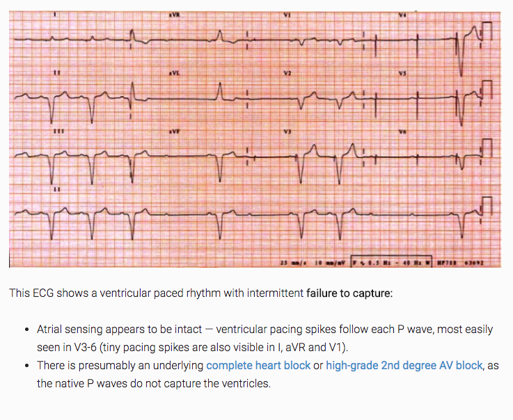
1. Failure to Capture- Pulse sent but myocardium doesn’t feel it.  No depolarization.  If myocardium is in refractory state, depolarization won’t happen. 1.Causes include electrode displacement, wire fracture, electrolyte disturbance, medications, heart disease, MI, or exit block.

2.N.B.  If native rate above pacemaker threshold, it naturally won’t send a signal.  That’s not failure to capture.

3. from LITFL!

2. Failure to Pace- Pacemaker not sending a pulse to stimulate the heart.  HR found below the lower limit set on the device and no pacemaker artifact on EKG.

1.Pacemaker over-sensing is most common cause of failure to pace.

2.Lead fracture from blunt trauma due to MVA, sports, or falls

3.Over-sensing caused by device misinterpretation of myopotentials from surrounding muscle (e.g. someone painting a room, causing contraction of the pectoris).  Can also be due to retrograde p waves, t waves, or other post-depolarization electrical activity sensing as though it was a normal potential and causing the pacemaker not to send a stimulus.

4.OVER SENSE=FAILURE TO PACE

5.May see large P or T waves, skeletal muscle activity on EKG or may not see anything.  If patient stimulates rectus or pectoral muscle while on cardiac monitor, may see reduced pacemaker output.

3. Failure to Sense- Pacemaker doesn’t recognize a myocardial depolarization (native activity) after it’s traveled up the lead wire.

1.“Blanking” where the pacer doesn’t sense a normal depolarization.  Prolonged refractory period can be 2/2 changes in the patient’s EKG complex (e.g. new BBB) and cause functional UNDER-sensing.  Usually lead fracture or insulation defects.

1.May see pacing spikes WITHIN the QRS complex.

2.Get asyncronous pacing

3.UNDER SENSE = FAILURE TO SENSE

2.Causes:  increased stimulation threshold at electrode site (exit block), poor lead contact/fracture/insulation defects, new BBB or programming problems

http://www.emdocs.net/pacemaker-and-aicd-management-in-the-emergency-department/

Rosen’s Chapter 70:  Implantable Cardiac Devices

https://lifeinthefastlane.com/ecg-library/pacemaker-malfunction/

 ·