POTD: Uric Acid in Urinalysis

POTD: Uric Acid in Urinalysis

What do you make of this “moderate uric acid” in this UA?

ur2.JPG
ur1.JPG

I usually just overlooked it whenever I saw it on a UA…I’m sure its fine…but now that I’m on admin, I got a bit of time to take a closer look. For the most part, it doesn’t really mean anything, but there are cases where it can assist in our decision making.

Purine nucleotide (sardines, veal, in basically every proteinaceous food) metabolism produces relatively water-insoluble uric acid in humans. Uric acid level is determined by rate of synthesis, rate of renal/GI excretion, and metabolism. Uric acid has a pKa1 of 5.3 so it’s water soluble at physiologic pH of 7.4. Urine has a wide range of physiologic pH 4-8 and as it gets close to 5.5, it starts precipitating from urine forming crystals. 

By far the most common cause of low urine pH is dehydration especially if the patient is having diarrhea which causes a loss of sodium bicarbonate (hyperchloremic acidosis). This can cause a drop in urine pH beyond 6 into the 5s which will lead to precipitation of uric acid crystals. The patient just needs rehydration and the urine pH will increase resolving the uric acid crystals. Patients can also develop uric acid crystals if their diet is too protein rich so dietary modification can be indicated.  This is why patients with many gout flare ups are often on low purine diets.

Uric acid stones make up 5-10% of all kidney stones in the US.  The thing is that in the most common calcium oxalate stones, there is often urate excreted as well so sometimes uric crystals are not always diagnostic. Uric acid crystals are radiolucent on XR but easily visible on non-contrast CT, but who is trying to find stones with an XR anyway? The treatment is just hydration and at most diet modification.  It’s interesting that in desert/arid regions of the world, up to 40% of all renal stone are due to uric stones.  The patient might have a condition causing hyperuricemia leading to recurrent uric acid stones (gout, malignancies, hemolytic disorders, obesity, G6PD, purine overproduction genetic disorder).  The next step would be alkalization of the urine; most commonly daily potassium bicarbonate or potassium citrate can be given to dissolve the stones, but this is not really an ED concern. PMDs/urologists can do serum and urine uric acid testing if these stones do not resolve to identify if the hyperuricemia is from overproduction or underexcretion.  I could not find a study finding the correlation between uric acid crystals and the likelihood of kidney stones.  There are studies linking lower pH to increased chance of kidney stones but there were no specific numbers given.

One of the patient populations where uric acid crystals is more concern are cancer patients are chemotherapy.  Chemotherapy/radiation can induce rapid cell lysis leading to hyperuricemia in tumor lysis syndrome. This can lead to acute uric acid nephropathy when uric acid precipitate on the nephrons causing acute uric acid nephropathy. Sometimes these patients need to be on rasburicase, allopurinol, or febuxostat to reduce uric acid production. The problem with uric acid crystals in urine is that it can be negative this in case if they become obstructed in the nephrons.  It does not always correlate with uric acid level, which is a much better indicator of hyperuricemia. Indication for dialysis in tumor lysis syndrome is serum uric level > 10, renal failure.

Gout is characterized by extracellular fluid urate saturation leading to hyperuricemia.  These patients have 2x likelihood of having kidney stones but the most common stone is still calcium oxalate. In these patient’s uric crystal in urine is still not correlated to uric acid level. Gout is diagnosed with arthrocentesis of the synovial fluid. Urine uric crystals are not useful in diagnosis in this case as the uric acid is precipitated in an acute attack and the patient’s uric acid levels are often (70%) normal. Serum uric acid levels are not even very helpful in these cases.

Overall, seeing uric acid crystals in urine is generally just a sign of urine pH < 6 likely due to dehydration. It can help guide the management by the urologist or primary care doctor after the visit but it is of low utility to the ED provider.  It can tip us off for tumor lysis syndrome or a gout attack, but I wouldn’t depend on it.  Serum uric acid level are much more useful from a diagnostic perspective.


POTD: Arrhythmogenic Right Ventricular Dysplasia/Cardiomyopathy (ARVC)

POTD: Arrhythmogenic Right Ventricular Dysplasia/Cardiomyopathy (ARVC)

Case: 22M presents unconscious in VTach.  The patient was exercising and suddenly passed out.  ACLS performed and after defibrillation, ROSC was achieved. Epsilon wave was seen on EKG, pt was admitted to MICU, which did MRI showing arrhythmogenic RV dysplasia.

Arrhythmogenic Right Ventricular dysplasia:

Inherited myocardial disease where R ventricular myocardium is replaced by fibro-fatty tissue

Typically autosomal dominant but there is recessive form caused Naxos Disease with wooly hair and skin changes

3:1 men to women, more in Italian or Greek descent, 1:5000 people overall

Second most common cause of sudden cardiac death to HOCM (causes 20% of sudden cardiac death in pt < 35yo)

Presentation: 30-50s with syncope or dysrhythmias including PVCs, VT, VF, CHF, or sudden cardiac death

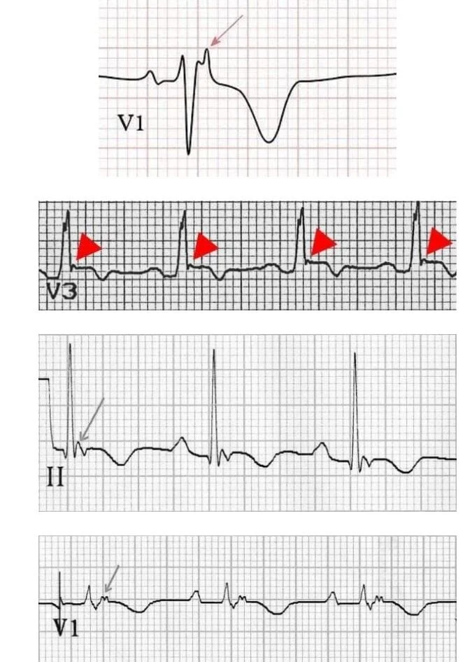
epsilon wave.JPG
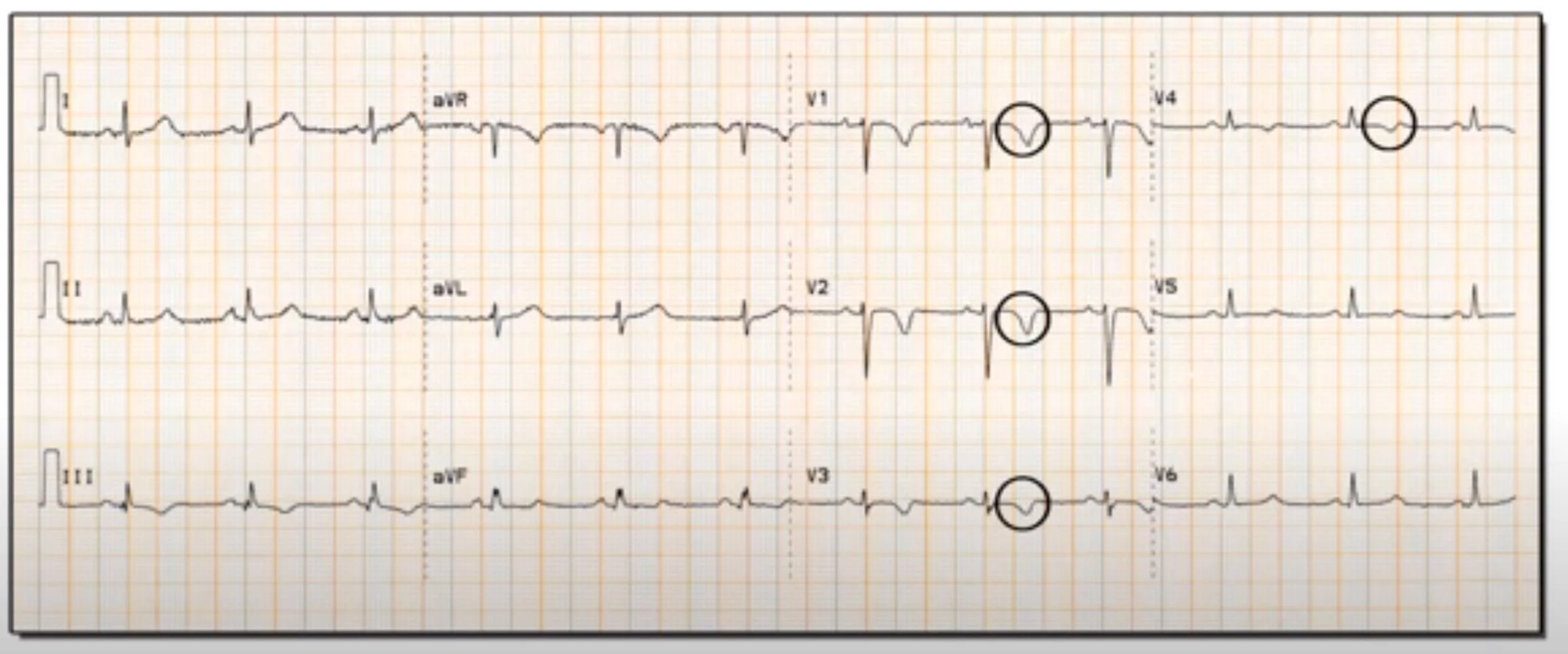
TWI.JPG
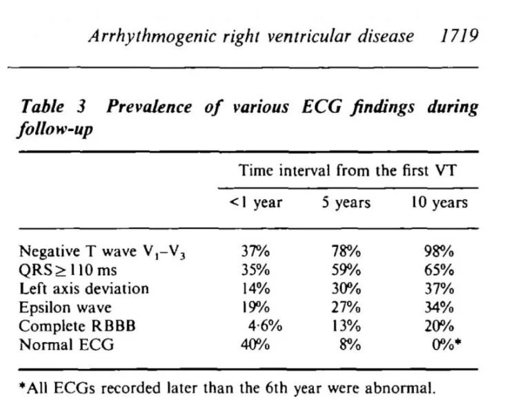
ekg resulst.JPG

EKG:

epi1.JPG

Epsilon waves are the most specific finding on EKG (30-50% spec) but there are other findings as well, the most common being TWI in V1-V3. The epsilon wave occurs because conduction is slower through fattier tissue, and there is a post-excitation wave. 

It can be difficult to pick up epsilon waves, a different set of lead placement can be used

RA over manubrium, LA over xiphoid, LL in V4 position, I/II/III will be denoted as FI/II/III in the image

Fountain.JPG

Imaging:

Echo – dilated hypokinetic RV with prominent apical trabeculae and dilated RV outflow tract

MRI – will see fibrofatty infiltration, RV dilation, wall motion abnormalities

Treatment:

Betablockers preferably Sotalol or amiodarone

ICD placement if patient has sypmtoms, some pathways can be ablated

Some patients will develop heart failure and will require standard CHF treatment, other might need heart transplant

Disposition:

If patient is asymptomatic and abnormal EKG is found incidentally, the patient can be discharged with cardiology f/u

The patient should be admitted if concerning symptoms likely syncope, chest pain, arrhythmias or has a family history of sudden death/cardiac arrest


POTD: Brugada Syndrome

POTD: Brugada syndrome

Brugada syndrome is a cardiac disease cause by an inherited ion channelopathy (Na, K, and Ca channels affected) with a propensity to develop Vfib, Vtach, Afib, and rarely SVT leading to sudden death. Most events happen between midnight to 6am - in Thailand, Brugada Syndrome is known as "Lai Tai" or "death during sleep".

Demographics: asian, average age 41 yo, males are 9x more likely

EKG can be sinus then Brugada can triggered by fever, vagal tone, cocaine/alcohol use, ischemia, hypoK, hypothermia, medications (sodium channel blockers, calcium channel blockers, nitrates, a agonist, B blocker)

History: ask for family history of sudden death < 45yo, syncope and cardiac symptoms in the past

EKG look in V1/2

Brugada.jpg

Type 1: elevated ST> 2mm that descends with upward convexity to TWI

Type 2: elevated ST> 1mm that descends toward baseline then rises again to upright T wave

Type 3: elevated ST> 1mm descends toward baseline then rises again to upright T wave


Treatment

If patient is asymptomatic and Brugada EKG was found incidentally, you can discharge with cardiology follow up.

If the patient has syncope, chest pain, sob or any concerning symptoms, admit for EPS study and ICD placement.

If the patient is in tachyarrhythmia (Vfib, Vtach, etc) and the patient is unstable, cardiovert

If the patient is stable, requiring recurrent cardioversion, or having recurrent shocks from pt's ICD (people known to have 50+ shocks etc.), you should treat medically. Ideally, consult cardiology for recommendations. If you are solo coverage in community without cardiology...oh God...

Given the channelopathies, many commonly used VTach medications for Brugada are contraindicated or ineffective.

www.brugadadrugs.org is a great site to tell you which medications should not be used: procainamide, flecanide, bupivacaine, propofol, ketamine, tramadol, lithium

Amiodarone is ineffective, case studies have found that it can unmask hidden Brugada but will not terminate the rhythm. BB and CCBs are also shown to be ineffective.

Quinidine is the best studied medication for tachyarrhythmias in Brugada, terminating 80% of arrhythmias. (Interestingly it is a Na channel blocker, but it's blockade effect on Ito an outflowing K channel is what helps it treat Brugada)

Immediate release form 200-400 mg PO q6h, then extended release 324 mg PO q12

Isoproterenol (B1/B2 agonist) is less studied but it can be given as a drip at 2-10 mcg/min tritrated to effect.

I spoke to pharmacy at Maimo and they were kinda enough to ask Dr. Adzic who said he would use Quinidine or Isoproterenol not amio.