Physician Mortgage Loans

What is a physician mortgage loan?


A physician loan is a mortgage loan that requires the borrower to have a DM, DO, DPM, DVM, DDS, or DMD degree (there may be more acceptable degrees than that depending on location and bank). Most of these loans typically require you are still in training or within ten years of completing training. This type of loan package enables medical professionals to effectively borrow more money than they otherwise would with a conventional mortgage loan. These loans can be up to $1,000,000! 

What is the benefit of a physician mortgage loan?

One significant benefit of physician mortgage loans is the ability to purchase a home without the requirement of a down payment. This is advantageous for medical professionals who may have substantial student loan debt and limited savings. Additionally, these loans often do not mandate private mortgage insurance (PMI), which is typically required when the down payment is less than 20% on a conventional mortgage. By eliminating the need for PMI, physicians can potentially save on monthly mortgage costs. The calculation of the debt-to-income (DTI) ratio, a crucial factor in mortgage approval, is approached differently for physician mortgage loans. Lenders exclude certain debts, such as student loans, from the DTI calculation, making it easier for doctors to qualify for a larger loan amount.


Are there any drawbacks to a physician mortgage loan?

Interest rates on physician mortgage loans may differ from those of standard mortgages or first-time homeowner loans. Often they will be slightly worse than conventional mortgages. The difference is typically not substantial, but often the recommendation may be to refinance the mortgage after a few years to get a better interest rate. This will be situation dependent. Also note, not all banks offer these types of loans.

TLDR: You can qualify for a $1,000,000 mortgage loan with no down payment, and no PMI, if you are in medical training or within ten years of completing training. 


Spider Bites

The brown recluse spider, scientifically known as Loxosceles reclusa, is known for its venomous bite. This spider is found in southern US states.

  1. Mechanism of Toxin: Sphingomyelinase D causes hemolysis and complement mediated erythrocyte destruction. There are multiple proteases that break down collagen, elastin, fibrinogen, etc and act synergistically with sphingomyelinase D to cause local tissue destruction

  2. Clinical Features of Bites:

    • Course of bite: The bite is often painless or with minimal pain. There will be two small puncture wounds. This will become pale with the edges becoming red. Over the next few days, this turns into a blister with a central ulcer, followed by skin sloughing. Can take weeks for wound to heal.

    • Early Symptoms (2-8 hours):

      • Redness and swelling around the bite site

      • Mild to moderate pain and itching

    • Delayed Symptoms (12-36 hours):

      • Necrotic (dead) tissue formation, leading to an ulcer

      • Systemic symptoms like fever, chills, malaise, headache, nausea

    • Worsening complications:

      • DIC

      • Rhabdo

      • Kidney Failure

  3. Evaluation: Lab tests should only be ordered in patients with systemic symptoms and fear of worsening complications. Should order CBC, CMP, CK, retic count, haptoglobin, LDH, PT/INR, D-dimer, fibrinogen.

  4. Medical Management:

    • Local Wound Care: Primary management is local wound care. Clean the site with soap and water, apply a cold compress to reduce swelling, and elevated the affected site. Sphingomyelinase D also has reduced activity in lower temp, so ice packs are important! Patient should also receive tetanus prophylaxis!

    • Pain Management: NSAIDs

    • Wound Care for Necrotic Tissue: If wound is severe enough, may require eval for debridement and potential skin grafting (this is usually weeks later). Hyperbaric oxygen therapy can also be considered for severe cases.

    • Antibiotics: Only if concern for local cellulitis.

    • Systemic Treatments: Weak evidence for use of dapsone. There is slightly more evidence behind the use of corticosteroids for reducing the risk of AKI and rhabdo.

It's crucial to note that brown recluse spider bites are rare, and most cases resolve with local wound care.


Black widow spiders, known as Lactrodectus spp, are venomous arachnids found in various regions around the world. The venom they produce contains neurotoxins, primarily alpha-latrotoxin, which affects the nervous system. These spiders classically have the “red hourglass” marking on them.

1.       Mechanism of Toxin: The primary toxin in black widow spider venom is alpha-latrotoxin. It works by binding presynaptic neurons, creating calcium permeable channels in the lipid layers, causing an influx of calcium into the presynaptic cells. This leads to an excessive release of neurotransmitters. Primarily concerned with release of acetylcholine.


2.       Clinical Features of Bites:

  • Course of bite: Bites are often initially characterized by severe local pain at the bite site. Very quickly patients will develop erythema and edema at site of bite

  • Systemic Symptoms: As the venom spreads, systemic symptoms may develop, including muscle pain and cramps, abdominal pain, weakness, sweating, and nausea. Patients may experience autonomic nervous system effects such as increased blood pressure and heart rate.

  • Worsening complications

    • Rhabdo

    • Myocarditis

    • A-fib

3.     Laboratory Tests: Lab values are generally nonspecific for black widow bites. Patients will tend to have elevated WBC, hematuria, and elevated liver enzymes. There are documented cases of rhabo and myocarditis from black widow bites, and there for kidney function and troponins can be checked if patients complain of systemic symptoms.

4.       Medical Management:

  • Local Wound Care: Clean the site with soap and water. Patient should also receive tetanus prophylaxis!

  • Pain Control: Analgesics, such as opioids or muscle relaxants, may be used to manage pain.

  • Antivenom: In severe cases or when systemic symptoms are significant, antivenom may be administered. This can rapidly reverse the effects of the venom. It is horse derived, and may cause anaphylaxis.

  • Observation: Patients may be observed for several hours to ensure symptoms do not worsen and to monitor for potential complications. Consider admission in children, patients with preexisting cardiac conditions, pregnant women, or for severe symptoms.

    It's important to note that while black widow spider bites can be painful and cause distressing symptoms, fatalities are rare.


    Anoka IA, Robb EL, Baker MB. Brown Recluse Spider Toxicity. [Updated 2023 Aug 7]. In: StatPearls [Internet]. Treasure Island (FL): StatPearls Publishing; 2024 Jan-. Available from: https://www.ncbi.nlm.nih.gov/books/NBK537045/

    Williams M, Sehgal N, Nappe TM. Black Widow Spider Toxicity. [Updated 2023 Aug 7]. In: StatPearls [Internet]. Treasure Island (FL): StatPearls Publishing; 2024 Jan-. Available from: https://www.ncbi.nlm.nih.gov/books/NBK499987/


VOTW: Regional Wall Motion Abnormality

This weeks VOTW is brought to you by Dr. Eng and Dr. Xu!

An 82 year old male presented to the ED w/ confusion, slurred speech and fall. A stroke alert was called initially, however the EKG obtained showed deep inverted T-waves in the anterior leads as well as ST-elevation in I and aVL. A POCUS was performed which showed…

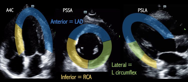
Clip 1 shows a parasternal short axis view of the heart. The septum, posterior and inferior walls appear to be contracting appropriately but the anterior and lateral walls appear akinetic. Clip 2 shows an apical 4 chamber view of the heart where again the septum appears to be contracting well but the apex and lateral walls appear to be akinetic. The area of akinesis correlates with the ST-changes seen on the EKG.

SALPI

Regional Wall Motion Abnormality

To evaluate for a regional wall motion abnormality (RWMA) remember the acronym SALPI (image 1). In the parasternal short axis view, starting at the septum, go clockwise to identify the anteriorlateralposteriorinferior walls. To look for a RWMA, look closely at each wall during systole to see if:

  1. The myocardium is moving in towards the center of the ventricle

  2. The myocardium is increasing in thickness

The absence of these findings is concerning for a RWMA which may be indicative of an acute MI. Patients with old MIs may also have RWMAs - correlate with the EKG and old echos if available

The parasternal long axis view and apical 4 chamber views can also be used to evaluated for RWMA (image 2).

When to POCUS for RWMA

This may be especially helpful in patients w/ equivocal EKGs that you or cardiology is on the fence about activating the cath lab or when the symptoms are not quite consistent with an MI (as in this case). Finding a RWMA may expedite cath lab activation (1).

Pro Tip: Cover up the entire LV with your hand except the specific wall you’re looking at and look at each wall seperately.

Back to the patient

The patient did not have any active chest pain but the initial troponin returned at 27.

The patient was taken to the cath lab which showed triple vessel disease with 80% stenosis of mid-LAD, 95% stenosis of first diagonal, 95% stenosis of proximal circumflex. He was evaluated for CABG but ultimately chose medical therapy.

References:

(1) Xu C, Melendez A, Nguyen T, Ellenberg J, Anand A, Delgado J, et al. Point-of-care ultrasound may expedite diagnosis and revascularization of occult occlusive myocardial infarction. Am J Emerg Med. 2022;58:186–91.