Ocular Ultrasound

For today's POTD, we'll be taking a closer l👀k at eye pathology, specifically how we can use ocular ultrasound to further evaluate many common eye complaints encountered in the ED.

While eye complaints are fairly common in the ED, our comfort level with the diagnostic tools available to us for these complaints varies greatly.
These tools include:
- Slit lamp (the hardest part is really finding and flipping the 4 separate switches to actually turn it on)
- Fluorescein and Wood's lamp
- Tonopen (addressed in a previous POTD)
- Bedside fundoscopy (I'll admit I suck at this)
- And, ocular ultrasound!
The biggest advantage of ocular sono is how easy it is to perform.
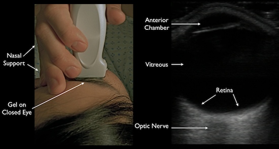
1) Slap on a tegaderm over the patient's closed eye
2) Squeeze on a generous amount of ultrasound jelly
3) Gently lay on your linear probe and look both horizontally and vertically (tell the patient to look straight ahead behind their closed eyes)

You can make so many diagnoses using ocular ultrasound!!!
- Foreign body
- Globe rupture
- Lens dislocation
- Retinal detachment
- Vitreous detachment
- Increased ICP (seen through increased optic nerve sheath diameter: measure 3 mm down, then 5 mm across)

We'll quickly focus in on retinal vs vitreous detachments. This is explained wonderfully in video form at http://5minsono.com/rdvd/, which I will summarize here.


In a retinal detachment, you can see the lines connectingto the optic nerve*. [How to remember this: the retina is just an extension of the optic nerve]

*To get a little more technical, you can also use ultrasound to tell whether a retinal detachment is "mac-on"(macula still attached, i.e. need for emergent surgical reattachment) or "mac-off" (macula detached, i.e. damage probably already done).
- The key to understanding this is knowing that the macula is located lateral to the optic nerve (or in eye-speak, on the temporal side).
- This 1 minute video illustrates the difference better than I can in words: https://www.youtube.com/watch?v=JijIfSzOG9U
- I think in reality though, if I suspect any kind of retinal detachment in a patient, whether it's mac-on or mac-off, I'll be transferring them for emergent ophtho evaluation.


vitreous detachment is typically seen floating above and NOT connected to the optic nerve. It is often also hazier and swirlier (very technical terms), especially as the patient looks in different directions.

References:
Academic Life in EM
5MinSono.com
EMin5.com
 · 

baby its cold outside...

Today's POTD inspired by two similar yet very different resus patients this morning...So lets talk HYPOTHERMIA.. tis the season!

Definition/Classification:

Causes: AGE IS BIGGEST RISK FACTOR- THINK ELDERLY AND INFANTS!
  • Heat loss- environmental exposure, vasodilation
  • Decreased or impaired thermogenesis:
Endocrine
Hypothyroid, adrenal insufficiency, malnourishment
CNS
Hypothalamic lesions, hypopituitarism
Trauma
Burn, spinal cord injuries
Sepsis
 
Tox
 ETOH, sedatives, vasodilators
Skin
Psoriasis, exfoliating conditions
Psych
 
Iatrogenic
Cold fluids, intraop
Evaluation:
  • Get that probe in! Core temperature is actually better obtained from esophageal or bladder > rectal
  • BGM!
  • Labs: Coags, CBC, BMP, CPK, TSH, sepsis workup?
  • EKG: Osborn waves, T wave inversions, prolongation of PR/QRS/QT, AV block, PVC
    • VF highest when T, 28C
Management: Think about and treat causes!

  • Passive Warming: Mild- think pts able to shiver
    • Warm blankets, keep dry, insulation
  • Active Warming
    • External/Peripheral- Bairhugger, warm fluids,chemical heat pads ( watch for burns)
      • May have drop in temperature from cold diuresis and release of cold peripheral blood to core
    • Central active warming
      • Warmed (40-46C) humidified inspired gases (1 C/h; 1.5°C/h ET tube)
      • Warm IV fluids (42C)
      • Body cavity lavage with 40C fluid e.g. peritoneal (3C/h), gastric, bladder, right-sided thoracic lavage (3-6C/h – use 2 ICCs for continuous flow)
      • CRRT
      • ECMO/ bypass (9-18C/h)
  • Coding:
    • Gentle handling of patient- can cause VF!
    • Cold heart is resistant to meds and pacing hold until temp >30C
    • Double the dose when temp bwn 30-35C
    • Shock Resistant- sources say shock max of 1-3times, goal is to get them warm!
Complications: Acid base disorders, pneumonia, bleeding, other cold injuries, dysrhythmias, DIC, rhabdomyolysis, thromboembolism, pancreatitis, multiorgan failure. Hope everyone is staying warm outside, enjoy the snow


sources: life in the fast lane, emdocs

 · 

PE in Pregnancy

Welcome to our exciting Thursday edition of POTD, in which we will be talking about...

PE in pregnancy!
Special thanks to Drs. Hoke and Koch for the inspiration. 

We all learn in medical school that pregnancy is a hypercoagulable state, and that these expectant moms are at increased risk of PE and DVT because of this. But to what extent should we be fearing this potentially deadly diagnosis in the pregnant patient who complains of shortness of breath?
The idea that pregnant patients are at higher risk for PE came from epidemiological studies showing a 2-3 fold increase in DVT and PE during pregnancy. However, these studies did not differentiate between DVT and PE, and 2/3 of the cases were actually just DVTs -- does this change your perception?
In a 2015 meta-analysis by Meng et al, only 3 of every 10,000 pregnancies was found to have PE. The actual time of highest risk is POST-delivery, particularly in C-section patients, up to 1 month post-partum.
In a 15 min long EM:RAP segment from March 2016, Dr. Jeff Kline (the PE guru of the EM world) outlines his own reasonable approach to PE workup in our pregnant patients.
1) Start with bilateral lower extremity dopplers looking for DVT
2) If negative, can evaluate the patient using PERC criteria. If the patient PERCs out, discuss with mom either stopping the workup, or continuing on with a D-dimer.
3) Much has been said about trimester-adjusted D-dimer levels. While there are no validated studies on this, Kline proposes cutoffs of 750 ug/mL, 1000 ug/mL, and 1250 ug/mL for the first, second, and third trimesters (assuming an assay with usual cutoffs at 500 ug/mL). I believe our assay cuts off at 380, so not sure how these values figure comparatively...
4) If the D-dimer comes back elevated, the next step is to proceed with pulmonary vascular imaging.
Often, the mom may ask, "What's the actual risk of radiation to my baby?" The real answer is no one can be sure, but here are the fetal doses of radiation from common radiologic studies (from ACOG): 
- 2 view plain CXR: 0.0005 to 0.01 mGy (milliGray)
- CT pulmonary angio: 0.01 to 0.66 mGy
- Low dose V/Q scan: 0.1 to 0.6 mGy (perfusion-only scan delivers less radiation, too)
Generally speaking, a fetal radiation exposure of <5 mGy is considered a safe dose in pregnancy.
This chart shows the estimated upper limit radiation dose for teratogenic effects, which changes as the fetus develops:

ACOG recommends CT angio over V/Q scan to rule out PE in pregnancy. Why? 
- You can see above that a V/Q scan is actually a higher dose of radiation to the fetus. If for whatever reason you cannot get a CT angio, instruct mom to empty her bladder after the V/Q scan is completed, to minimize continued exposure of baby to the tagged radioisotope.
- Next, while CT angio has the added exposure of IV iodinated contrast which can cross the placenta, animal studies have not shown it to have any teratogenic effects.
- Lastly, breastfeeding women have traditionally been told to halt breastfeeding for 24 hr after getting IV iodinated contrast. This is a myth! ACOG states that <1% of iodinated contrast is excreted into the breast milk, and a further <1% of that <1% is actually absorbed through the infant’s gastrointestinal tract. So no real need to pause breastfeeding!
At the end of the day, your clinical gestalt is still key. Also utilize adjunctive tests (bedside echo looking for right heart strain, EKG, CXR) to guide your plan of action.
References:
EM:RAP
 ·