POTD: Superficial Thrombophlebitis (feat. Dr. Doge Pologe)

Today's POTD is inspired by resident extraordinaire Dr. Doge Pologe. As usual, TL;DR is below the main text. 

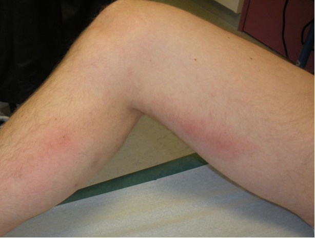
Thrombophlebitis is essentially a composite of two diagnoses: phlebitis, which is a clinical diagnosis in the setting of an erythema and pain overlying a vein and an identified thrombus. In the lower extremities, this is most likely to occur in varicose veins. 

1-Figure1-1.png

In general, risk factors for thrombophlebitis are the same as for DVT (think Virchow's Triad of hypercoagulability, hemodynamic changes (stasis/turbulence), and endothelial injury/dysfunction). These include pregnancy, history of vein excision/ablation, history of prior thrombosis, malignancy, and prior IV catheter placement. 

Special cases:

  • "Mondor": thrombophlebitis of a breast vein, anterior chest vein, or of the dorsal penile vein. The two former should prompt a search for breast cancer and the later is usually due to repetitive trauma

  • "Trousseau's sign of malignancy": migratory thromboembolism, has a strong association with adenocarcinoma of the pancreas and lung


Diagnosis
:
Diagnosis of phlebitis is clinical. Ultrasound should be used to identify a thrombus to distinguish between phlebitis and thrombophlebitis. Patients should also have duplex ultrasound to identify a DVT especially if the area of concern is above the knee. This is important because the rate of concurrent DVT in all cases is 25% and the rate of concurrent PE is 5%. 

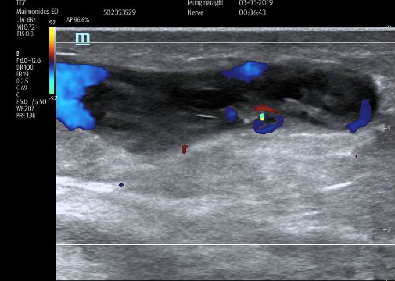
Thrombophlebitis on ultrasound will demonstrate heterogeneous internal echoes within a superficial vein. Unlike an abscess or a lymph node, this will not be discrete and you should be able to trace it out. in the words of the Doge:

"it looks like a weird continuous twisty spaghetti
abscess thing; 
but it's not to be feared, 
mostly superfluous, so get frisky and ultrasound that biddy, 
color doppler, diagnosis, ka-ching". 

There may be flow present (which can help distinguish an abscess from thrombophlebitis; abscess = no flow). Some examples (by the Doge Pologe and myself):

thrombophlebitis.png
Screen Shot 2019-04-15 at 6.57.02 PM.png

Treatment:
As with treatment of below the knee VTE, the treatment of superficial thrombophlebitis is controversial. Patients can be considered low risk (for VTE) if they meet these criteria:

  • Affected vein segment < 5cm

  • Remote from the saphenofemoral/saphenopopliteal junction

  • Low risk for VTE

**Repeat duplex should be obtained in 7-10 days or for worsening symptoms to check for propagation!

NB: thrombophlebitis post-ablation are always low risk and do not require treatment

For these uncomplicated cases, treatment is aimed at alleviating symptoms and prevention of propagation. This includes the following:

  • NSAIDs

  • Warm/cool compresses

  • Elevation of the extremity

  • Compression stockings

Patients that do not qualify as low risk or if they have recurrent thromboembolism should be considered for anticoagulation. Although studies looking at anticoagulation for SVT are small and flawed, NSAIDsLMWH, and fondaparinux have all been shown to decrease incidence of DVT. Patients can also be discharged on Xarelto (this is the only NOAC to be studied for this indication). 

In addition, thromboembolism can become suppurative. Signs and symptoms include high fever (as opposed to the low-grade fever that accompany simple thrombophlebitis) and purulent drainage (duh). In these cases, consider antibiotics


TL;DR

  • Thrombophlebitis = phlebitis (redness/pain along vein) + thrombus

  • Find the thrombus on ultrasound! Look for internal echoes.

  • Low risk patients = below the knee, affected vein < 5cm, distance remove from saphenofemoral/saphenopopliteal junction can be managed with NSAIDs, compression stockings, warm/cool compresses, elevation

  • Low risk patients should get repeat study in 7-10 days to check for propagation

  • High risk patients: consider anticoagulation with LWMH, fondaparinux, or Xarelto

  • Antibiotics for suppurative thrombophlebitis

Special thanks to Dr. Jonas Pologe, Dr. Lawrence Haines, and Dr. Leily Naraghi Bagher Pour. 

Sources:
http://www.emdocs.net/core-em-superficial-venous-thrombosis-svt/
https://www.bmj.com/rapid-response/2011/10/31/superficial-thrombophlebitis-not-straightforward-we-may-think
https://emedicine.medscape.com/article/463256-overview
https://www.uptodate.com/contents/phlebitis-and-thrombosis-of-the-superficial-lower-extremity-veins
https://radiopaedia.org/cases/superficial-thrombophlebitis-1?lang=us

 · 

Ultrasound-Guided PIV Placement Part 2 POD

The following is a powerful advanced technique that can be used to troubleshoot traditional short-axis US-PIV placement.

A familiar scenario: The elusive needle tip!

  • You’re placing an US-guided PIV and going ahead with your short axis technique

  • The vessel is directly under the center of the probe, right on that Bx-guide line

  • You know exactly how deep it is

  • The needle has entered the skin… the tip should be right over or very near the vessel…

  • But where is it??? You’re bouncing the needle a little and see tissue moving, you’re slowly sweeping the probe backward and forward where your needle tip should be, but it continues to elude you! Maybe it’s a little deep, maybe there’s some echogenic (bright) tissue hiding it, doesn’t matter, here’s what to do...

The answer: Long Axis — Hear me out!

  • Take your eyes off of the ultrasound screen

  • Pick up the probe and place it back down, marker toward you, exactly along the axis of the angiocath, DIRECTLY over it!

  • Without moving your hand, look back up at the screen. Unless you’re at a crazy steep angle, you will see your whole needle clearly!

  • If you see the vessel on the screen as well, you are now perfectly set up to continue placing your IV

  • Position the tip in the vessel lumen, then advance the angiocath over the needle as you normally would

Image result for long axis ultrasound guided IV

The other scenario: You’ve positioned the probe over the needle, you look up and see the needle but not the vessel any more, or maybe part of the vessel — here’s what you do

  • Slide/rotate the probe such that you have the vessel in view at its widest diameter on the screen

  • Then LOOK BACK AT THE ARM

  • If the probe is now to the right of the needle, you need to redirect to the right; if the probe is to the left, the needle needs to go left

  • Withdraw the needle a few mm and then redirect so that it is inline with the ultrasound

  • As you do this, look back up at the screen and you should see the needle coming into view

In a nutshell: If you’ve lost your needle tip

1. Use the probe to show you where the needle is

2. Use the probe to show you where the vessel is

3. With the probe over the vessel, position the needle so that it’s directly under the probe

4. Now all three are lined up and you’re ready to position the needle tip in the vessel lumen

A few last tips:

  • You can fine-tune your left-right control of needle tip in long axis by just moving the needle slightly one way or the other and seeing if it comes more into view or less into view — this will start happening automatically if you practice this technique a few times

  • I still recommend letting go of the probe and advancing the angiocath with non-dominant hand, however if an assistant takes the probe when you are ready to advance the angiocath, you can watch it go into the vessel and ensure that it is advancing smoothly into the lumen.

  • You can do this with one person as well but this requires advancing the angiocath and stabilizing the needle with one hand, which is more difficult and gives little tactile feedback as to whether it is advancing smoothly or meeting resistance

  • Once you’re comfortable with this long-axis technique, try doing the entire procedure in long axis. This tends to work very well for deeper, straighter veins.

  • There’s no reason you can’t switch back to short once you’ve found your needle tip and repositioned it; perhaps it’s a twisty vessel with multiple turns and you need to walk it in a little more - short axis is better for navigating in the left-right direction (as long as you’ve located your needle tip!)

  • Remember the concept of "angle of insonation": the steeper your needle angle, the more difficult it will be to see your needle because fewer ultrasound beams are bouncing back to the probe (more are being deflected in a different direction)

Jonas Pologe, PGY3, Emergency Medicine, Maimonides Medical Center

 · 

POD Aortic Dissection

A patient came to the north side today with an acute aortic dissection. Here are images obtained by the ultrasound team when the patient first came in.

A suprasternal view showing an intimal flap:

suprasternal.jpg

A short axis view of the abdominal aorta showing an intimal flap

abdominal.jpg

Diagnosis was made, BP meds started, cardiothoracic consulted, and CT expedited.

CT showed a severe type B thoracoabdominal aortic dissection:

CT-aorta.jpg

Aortic Dissection

Pathophysiology:

Tear in the intima (inner most layer), bleeding into the media (middle layer)

Pathophysiology_Theaorticdissectionsoriginatewithanintimaltearin_Ascendingaorta65Aorticarch10.jpg

Diagnosis of aortic dissection is very time sensitive:

mortality is directly proportional to time elapsed between symptom onset and diagnosis/treatment

.

How does it eventually kill you? (I think it’s important to ask this question about all disease processes)

acute aortic regurgitation —> cardiogenic shock

Cardiac tamponade —> obstructive shock

Major brach-vessel obstruction —> vasodilatory shock from dead organ or limb

Aortic rupture —> hemorrhagic shock

2 types that we care about: Stanford Type A and Stanford Type B

types.jpg

Type A

:

involves ascending aorta

— surgical — a/w aortic rupture, tamponade, aortic regurg, AMI, stroke — more common (68%)

Type B

:

does not involve ascending aorta — medical (BP control and monitoring) — a/w limb/organ ischemia  — less common, (32%) — usually originates just distal to L subclavian artery

Classic history: old person,

very hypertensive

;

abrupt onset

,

tearing/ripping chest pain

,

radiating to bac

k; a/w neuro symptoms e.g.

weakness/numbness

(due to vessel branch occlusion); a/w syncope/diaphoresis/N/V

Other risk factors include Marfan’s, connective tissue disease, FHx aortic disease, known aortic valve disease, recent aortic manipulation (e.g. TAVR, surgery), known thoracic aortic aneurysm, tobacco;  rarely 3rd trimester pregnancy, TB, syphilis,  vasculitis, blunt trauma

Classic physical: Pulse deficit (present in <20% of cases), unequal BP in upper/lower extremities, neuro deficits, signs of tamponade

Diagnosis:

Labs: basics, coags, trop, consider d-dimer (actually high sensitivity/NPV for dissection due to blood often clotting I false lumen)

CT angio aorta: gold standard for diagnosis of aortic dissection

CXR: not sensitive, not specific — sometimes mediastinal or aortic knob widening, few other nonspecific signs

TEE: is an excellent modality that’s in the works but we don’t have it operational yet

TTE: next best thing, as usual with ultrasound it’s specific but not sensitive - see below

Ultrasound for aortic dissection — obtain the following views:

Subxiphoid: look for pericardial effusion

Image result for subxiphoid effusion'

Parasternal long: look for effusion, look at the descending aorta, look for aortic regurg with color doppler and measure the aortic root (nl <4cm) if you want to be fancy

para-long.jpg

Suprasternal window:

look for dissection flap (image from University of Maryland department of cardiology)

Probe above the the patient’s sternum pointed inferoposteriorly with probe marker to patient’s left (assuming cardiology convention)

SSNV.jpg

Abdominal aorta scan: look for dissection flap from diaphragm to iliacs, also measure diameter in short and long

Management (From the AAC/AHA aortic dissection guidelines):

ACC AHA AoD Treatment-Algorithm

Note: When blood pressure is intact, first bring heart rate with beta blockers, then control pain, then see if they need further BP control.

 ·