Metacarpal Fractures

Trauma Tuesday!Metacarpal Fractures

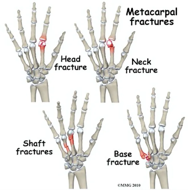
mcp fractures.png


Why do we care so much about a few small bones in the hand? Because missed injuries can lead to permanent disabilities--we (as well as our patients) need our hands for pretty much everything.

How to assess for these injuries? Do your typical hand exam but pay special attention to:

Rotational alignment! Have the patient flex at the MCP and PIP, forming a loose fist with the DIPs extended (as in the figure below, to the left). The axis of each digit should merge near mid wrist. Rotational malalignment will cause deviation of this axis for the injured digit.

mcp fractures 2.png


Rotational malalignment is usually an indication for operative repair, so be sure to check for it. 

Don't forget: Any open wound over the MCP should alert you to the possibility of a "fight bite"--usually require exploration or washout. This needs EMERGENT ortho evaluation. 

Diagnosis

Get X-rays - AP, lateral, and oblique views; pay special attention to the lateral as this is what you will use to measure angulation.

For the quick and dirty: acceptable shaft angulation is 40° for 5th MC, 30° for 4th MC, 20° for 3rd MCP, and 10° for 2nd. Reduce if there is greater angulation. 

Management

NONOPERATIVE: For stable fractures, those without rotational deformities, and those with acceptable angulation and shortening (usually 2-5mm for each shaft) => nonoperative repair: 

Reduce a dorsally angulated neck fracture before splinting, usually done via the Jahss technique. (https://youtu.be/40irKoUJqsM)

For MCP head/neck/shaft fractures, radial or ulnar gutter splint depending on which MCP is injured. For MCP base fractures, wrist splint. 


OPERATIVE: For open fractures, intra-articular fractures, fractures with rotational malalignment, significantly displaced or angulated fractures, or in the event of multiple MCP fractures => operative repair

Err on the side of prompt orthopedic follow up. 


Sources

https://emergencymedicinecases.com/episode-29-hand-emergencies/

https://coreem.net/core/metacarpal-fractures/

https://www.orthobullets.com/hand/6037/metacarpal-fractures

 · 

POTD Trauma Tuesdays: Fish Hook Removal

FISH HOOK REMOVAL

Introduction
▪ Most fishhooks consist of an eyelet at one end, a straight shank, and a curved portion that ends in a barbed point on the inner curve that points away from the hook’s tip. By design, it is constructed to prevent the hook from dislodging once it engages tissue
▪ Fish hooks are most often caught on hands and feet
▪ ED physicians may remove superficially embedded hooks but those embedded in vital structures (eyes, testicles, carotid artery, etc) should be referred to the appropriate surgical specialist covering that organ

How do I prepare to remove it?

▪ Stabilize the hook with a hemostat and remove any attachments, such as lures, fishing lines, sinkers, etc.
▪ Cleanse with betadine
▪ Use local anesthesia
▪ Children may need procedural sedation
▪ Pain control
▪ Tetanus prophylaxis

What methods are used for removal?
▪ Back out technique
⁃ If the hook is barbless, this is the easiest method.
⁃ As the name implies, back the hook out with a hemostat.

▪ Push through technique
⁃ Use when the tip of the hook is near the skin surface.
⁃ Push the hook through until you break the skin, and then use a wire cutter to cut the tip off.
⁃ Then back out the remainder of the hook.

▪ String technique
⁃ Hook’s belly should be directly in front of you with the shank pointing in the opposite direction
⁃ Loop a piece of string or large silk suture (3-0) around the belly of the hook and then wrap the ends around your index finger
⁃ Push down on the shank and eye of the hook with your other hand to disengage the barb from the surrounding tissue
⁃ Pull string slowly until it is taut in the plane of the hook’s long axis
⁃ Keeping it taut, jerk it quickly and firmly in the same direction

▪ Cut it out technique
⁃ When all else fails, cut with a scalpel along the hook, and then blunt dissect down with a hemostat.

Should I give antibiotics?

▪ No trials have investigated antibiotic therapy for fish hook injuries
▪ Most superficial fish hook wounds heal well without sequelae
▪ Consider antibiotics if the fish hook is deeply embedded in an infection-prone area such as a fingertip or ear
▪ Most infections are caused by skin flora
▪ If hook is contaminated (touched sea water, fish, bait, etc), consider abx treatment
⁃ Cephalexin 500mg PO q6 or cefazolin 1g IV q8 or Clinda 300mg PO q6 or 600mg IV q8
⁃ Seawater? ADD Doxycycline 100mg q12
⁃ See recent guidelines for other specific situations

 · 

Trauma in Pregnancy

Resuscitation of the Pregnant Trauma patient


General principles

·      Trauma is the most common cause of non-obstetrical maternal death in the United States

·      Best fetal resuscitation is good maternal resuscitation.

·      Stabilization of the pregnant women is the first priority; then, if the fetus is viable (≥ 23 weeks), fetal heart rate auscultation and fetal monitoring can be initiated and an obstetrical consultation obtained as soon as feasible

·      In Rh-negative pregnant trauma patients, quantification of maternal–fetal hemorrhage by tests such as Kleihauer-Betke should be done to determine the need for additional doses of anti-D immunoglobulin.

·      Tetanus vaccination is safe in pregnancy and should be given when indicated.


Airway

·      Greater risk for difficult intubation than non-pregnant patient

·      Pregnancy related changes à decreased functional residual capacity, reduced respiratory system compliance, increased airway resistance, and increased oxygen requirements

·      Gastric emptying is delayed in pregnancy à greater risk for aspiration

·      Respiratory tract mucosal edema à A smaller size of endotracheal tube is recommended

·      Choice of RSI medications NOT affected by pregnancy status


Breathing

·      Place chest tube one to 2 intercostal spaces higher than usual to account for displacement of the diaphragm during pregnancy

·      Marked increases in basal oxygen consumption à lower threshold for supplemental oxygen


Circulation

·      Fluid and Colloid resuscitation like standard trauma protocol

·      Uteroplacental vasculature is highly responsive to vasopressors, and their administration may decrease placental perfusion à vasopressors should be avoided unless refractory

·      Avoid supine hypotension: Compression of IVC by the uterus can cause up to 30% reduction in cardiac output à Place in left lateral position or by manual displacement of the uterus while the injured patient is secured in the supine position

·      O-negative blood should be transfused in order to avoid Rh sensitization in Rh-negative women

·      Vital signs: heart rate increases by 15% during pregnancy. Tachycardia and hypotension, typical of hypovolemic shock, may appear late in the pregnant trauma patient because of her increased blood volume.

·      Maternal vital signs and perfusion may be preserved at the expense of uteroplacental perfusion, delaying the occurrence of signs of hypovolemic shock

·      Attempt to obtain supra-diaphragmatic intravenous or intraosseous access for volume resuscitation and medication administration.


FAST

·      The FAST is less sensitive for free fluid in the pregnant patient than in non-pregnant patients.  Sensitivity decreases with increasing gestational age, likely due to altered fluid flow within the abdomen.

·      Management of suspected placental abruption should not be delayed pending confirmation by ultrasonography as ultrasound is not a sensitive tool for its diagnosis.


Secondary survey

·      In cases of vaginal bleeding at or after 23 weeks, speculum or digital vaginal examination should be deferred until placenta previa is excluded by a prior or current ultrasound scan.


Imaging

·      Radiographic studies indicated for maternal evaluation including abdominal computed tomography should not be deferred or delayed due to concerns regarding fetal exposure to radiation.

·      Ionizing radiation has the highest teratogenic potential during the period of organogenesis (5–10 weeks), with an increased risk of miscarriage before this period.

·      With abdominal CT during the third trimester the fetal exposure is around 3.5 rads, which is still under the threshold for fetal damage

·      Contrast agents should be used if indicated.


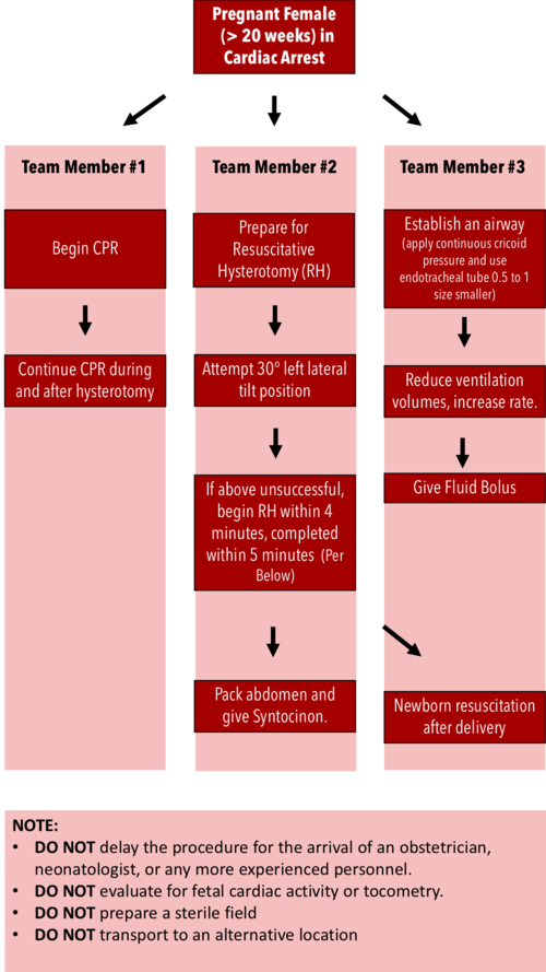
Resuscitative Hysterotomy in Cardiac Arrest

·      Should begin within 4 minutes and completed within 5 minutes of cardiac arrest

·      Both maternal and fetal survival decrease significantly after 5 minutes

·      Do NOT delay the procedure for the arrival of an obstetrician or neonatologist.

·      Do NOT evaluate for fetal cardiac activity or tocometry.

·      Do NOT prepare a sterile field (but be as clean as possible).

·      Do NOT transport to an alternative location.

·      Performing RH increases maternal cardiac output by 30%.


RH Algorithm.png


References:


Tamingthesru.com

EmDocs

Jain, Venu, et al. "Guidelines for the management of a pregnant trauma patient." Journal of Obstetrics and Gynaecology Canada 37.6 (2015): 553-571.

Smith, Kurt A., and Suzanne Bryce. "Trauma in the pregnant patient: an evidence-based approach to management." Emergency medicine practice 15.4 (2013): 1-18.


 ·