Frostbite

Frostbite is a condition that occurs when skin and underlying tissues freeze due to exposure to cold temperatures. It occurs due to vasoconstriction, resulting in decreased blood flow (which would deliver heat) to tissues, and this leads to ice crystal formation. Frostbite most often occurs due to exposure at temperatures of less than -20°C (that includes wind chill). Freezing alone typically does not cause tissue damage. Thawing is what worsens the damage by disrupting the endothelium of cells. The blood flow of skin is normally around 250 ml/min, but in tissue damaged by frostbite, drops to 20-50 ml/min. This endothelial damage leads to swelling, platelet aggregation, and potentially thrombosis (in the form of microthrombi due to decreased intravascular flow). 

Exposed and wet skin are the most likely to be damaged. Most likely areas are feet, hands, ears, lips, and nose. Risk factors for frostbite include patients that are colder temperatures, wind chill, no shelter, prolonged exposure, exposure while wet, patients with EtOH use. Patients with medical conditions that impact vasculature are also at increased risk, including diabetes, PVD, and smokers.


Classifications of Frostbite:

  1. First Degree: Involves superficial skin freezing, causing redness and irritation, but usually has good recover

    • Symptoms include stinging and burning, eventually throbbing

    • Patients typically develop erythema, swelling, and have desquamation later

  2. Second Degree: Involves freezing of the skin and deeper tissues, resulting in blistering and swelling, but usually has good recovery

    • Symptoms include numbness, then followed by aching and throbbing

    • Patients typically have extensive edema for hours, then develop blisters after that, and desquamate over the course of days

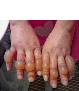
  3. Third Degree: Involves freezing extending to subcutaneous tissues, leading to deep tissue damage, usually has poor recovery

    • Symptoms include extremities feeling heavy, then burning, throbbing, and shooting pains

    • Patient with third degree frostbite will develop hemorrhagic blisters

  4. Fourth Degree: The most severe form, involving freezing of muscles, tendons, and bone, often leading to irreversible damage

    • Symptoms include a deep, aching pain

    • Skin becomes mottled, and deep eschar forms

Management:

  • Treat hypothermia first! Rewarm core temperature, otherwise at risk of reinjuring the patient if their extremities are re-exposed to cold again

  • Remove wet clothing

  • Place extremities in a 40-42°C bath for 20-30 min

    • Care should be made to not rewarm too quickly, otherwise you risk worsening reperfusion injuries

    • For areas that can not be placed in a bath (nose, ears, etc) can use a warm, wet towel

  • Rewarming is considerably painful and should be treated with aggressive pain control

  • Wound care should be undertaken

    • Wrap wounds with sterile, dry gauze

    • Keep effected extremities elevated

    • Do not remove blisters

    • Have a high clinical suspicion for possible compartment syndrome

  • Patients with full thickness injuries and no evidence of reperfusion after rewarming should be considered for tPa therapy which may reduce the risk of digital amputation

  • Patients should receive tetanus vaccination

Complications of frostbite

Patients can develop hypersensitivity to the cold with pain and ongoing numbness. Many patients can develop arthritis, can have loss of nails, cracked skin, atrophy of muscles. If tissue ischemia is severe, debridement may end up being necessary for extreme cases.


Example of Stage 3 injury

Basit H, Wallen TJ, Dudley C. Frostbite. [Updated 2023 Jun 26]. In: StatPearls [Internet]. Treasure Island (FL): StatPearls Publishing; 2023 Jan-Available from: https://www.ncbi.nlm.nih.gov/books/NBK536914/

Handford C, Thomas O, Imray CHE. Frostbite. Emerg Med Clin N Am. 2017;35(2):281–299.

Frostbite. Orthobullets. (n.d.). https://www.orthobullets.com/hand/12105/frostbite 

 · 
Share

Hypothermia

Hypothermia is a medical emergency characterized by a core body temperature below the normal range of 95°F (35°C).

Causes of Hypothermia:

  1. Increased heat loss

    • Homeless population

    • Elderly patients

    • Submersion injuries

    • Drugs, EtOH, CO poisoning can all cause increased vasodilation, leading to increased heat loss

  2. Decreased heat production

    • Endocrine (hypothyroidism, hypoadrenalism, hypoglycemia)

    • Erythrodermas (psoriasis, exfoliative dermatitis, eczema, burns)

    • Impaired shivering

    • Impaired thermoregulation

    • Sepsis

Swiss Hypothermia Staging System:

Stage 1: Mild (32-35°C) - Shivering, mild confusion, awake

Stage 2: Moderate (28-32°C) - Severe shivering, altered mental status

Stage 3: Severe (20-28°C) - Loss of consciousness, bradycardia, shivering may cease

Stage 4: Profound (<20°C) – Unobtainable vital signs

Associated Complications:

  1. Cardiac dysfunction

    1. Dysrhythmias can occur when body temperature drops below 30°C

    2. There is typically a drop in temperature and MAP after rewarming is started due to vasoconstriction

  2. Cold injuries (frostbite, etc. Maybe there will be more on this at a later date)

  3. Coagulopathy (patient may be coagulopathic despite normal labs because the lab rewarms the sample)

    1. Impaired clotting function

    2. Thromboembolism (due to hemoconcentration and poor circulation)

    3. DIC

  4. Impaired pharmacology

    1. Protein binding increases when temperature drops, rendering drugs ineffective

    2. Oral meds are not absorbed well due to decreased GI motility

    3. IM route is impaired due to vasoconstriction

  5. Rhabdomyolysis

General Management:

  1. Airway, Breathing, Circulation (ABCs)

    • Hypothermia causes a leftward shift in oxygen curve so support with oxygen, and prepare for intubation depending on how profound the hypothermia is

  2. ECG Findings

    • Patients usually have sinus bradycardia, can progress to a fib with slow ventricular response

    • Severe cases can develop v fib

    • Osborn or "J" waves (associated with moderate to severe hypothermia)

  3. Remove Wet Clothing - Prevent further heat loss

  4. Passive External Rewarming - Insulate the patient, provide warm blankets

  5. Active External Rewarming (should be done for moderate hypothermia)

    • Use forced warm air blankets or radiant heaters – our ED uses the Bair Hugger

  6. Active Internal Rewarming (for severe hypothermia)

    • Warmed intravenous fluids (warmed to 38-42°C)

    • Heated humidified oxygen

    • Various lavages (Thoracic, peritoneal, bladder, GI)

Management during Cardiac Arrest:

  1. CPR – initiate if patient does not have a pulse (should also assess if patient is still breathing)

    • It is challenging to assess vital signs in hypothermic patients - use end tidal or POCUS to help assist to see if patient is breathing and has cardiac function

    • Starting CPR if the patient does have a pulse may precipitate ventricular rhythms

    • Hypothermic patients have higher chances of improved neurological outcome and survival than normothermic patients that arrest

  2. Defibrillation

    • Use defibrillation if indicated, but note that hypothermic patients may not respond to defibrillation until adequately warmed

  3. ECMO

    • Patients with refractory hypothermia should be considered for ECMO

    • Patients with out-of-hospital-cardiac-arrest that are hypothermic should ideally be transported to an ECMO center

    • If patient is unstable (dysrhythmia, severe hypothermia, etc) ECMO teams should be contacted early in the ED visit


Stay warm out there this weekend!


Paal P, Pasquier M, Darocha T, Lechner R, Kosinski S, Wallner B, Zafren K, Brugger H. Accidental Hypothermia: 2021 Update. Int J Environ Res Public Health. 2022 Jan 3;19(1):501. doi: 10.3390/ijerph19010501. PMID: 35010760; PMCID: PMC8744717.

Baumgartner EA, Belson M, Rubin C, Patel M. Hypothermia and other cold-related morbidity emergency department visits: United States, 1995-2004. Wilderness Environ Med 2008;19:233-237

Brown et al., Accidental Hypothermia. N Engl J Med 2012; 367:1930-1938


Who to contact after a patient expires

I wanted to touch on a subject that is important but often not laid out in a concise manner - the protocol after a patient expires. 

I want to break this down into responsibilities of each of the staff. For residents (like myself) this often seems like a seamless process that happens in the background, but the reality is, multiple members of the ED are all coordinating together to progress this process.

Physicians- 

1. Attending physician must pronounce dead.

2. Admitting must be called with time of death, cause of death, whether or not the medical examiner will accept the case (more on that in a bit). Admitting will then process this info, and upload info to NYC Certify. The attending will then have to go into NYC Certify and certify the death.

3. The patient's family must be notified. Hopefully they are in the hospital, as it is more appropriate to have this conversation with the patient's family face to face, in private and to give them time to grieve with the patient.

4. Medical examiner must be notified in certain instances. The ME will take the following types of cases - trauma arrests, homicides, suicides, younger patients that are not terminally ill. Typically the ME will not take older patients with comorbidities. When in doubt, call the ME and they can decide.

5. Finally, the death note needs to be completed. 

Nursing- 

1. Charge nurse will call the expeditor / patient rep (more on that in a bit).

2. NYC LiveOn. This is the organ donor group. In our ED, nursing typically calls them. This requires answering questions about time of death, cause of death, medical comorbidities. 

3. Nursing and PCTs are typically responsible for post mortem care in patients that are not Jewish (more on the Guardians of the Sick in a bit). This involves removing lines, ET tubes, cleaning the patient, etc. This is NOT to be done in ME cases.

4. There is a written nursing protocol, on the MMC intranet site, I have shared the link below.

Expeditor/Patient Rep-

1. If the patient is Jewish, the expeditor will contact the operator, who contacts the Guardians of the Sick, who come and do post mortem care.

2. The patient rep may go to the family and offer support and comfort. Cannot provide suggestions regarding funeral homes (this is a conflict of interest).

3. Contact transport if the patient is going to our morgue (sometimes the family will arrange for the patient to be transported to a funeral home instead). 

Hope this helps outline the process and responsibilities during these stressful situations!

http://intranet.mmc/Main/DocumentLibrary/Post_Mortem_Care_2762.aspx

 · 
Share