Hypothermia

Hypothermia is a medical emergency characterized by a core body temperature below the normal range of 95°F (35°C).

Causes of Hypothermia:

  1. Increased heat loss

    • Homeless population

    • Elderly patients

    • Submersion injuries

    • Drugs, EtOH, CO poisoning can all cause increased vasodilation, leading to increased heat loss

  2. Decreased heat production

    • Endocrine (hypothyroidism, hypoadrenalism, hypoglycemia)

    • Erythrodermas (psoriasis, exfoliative dermatitis, eczema, burns)

    • Impaired shivering

    • Impaired thermoregulation

    • Sepsis

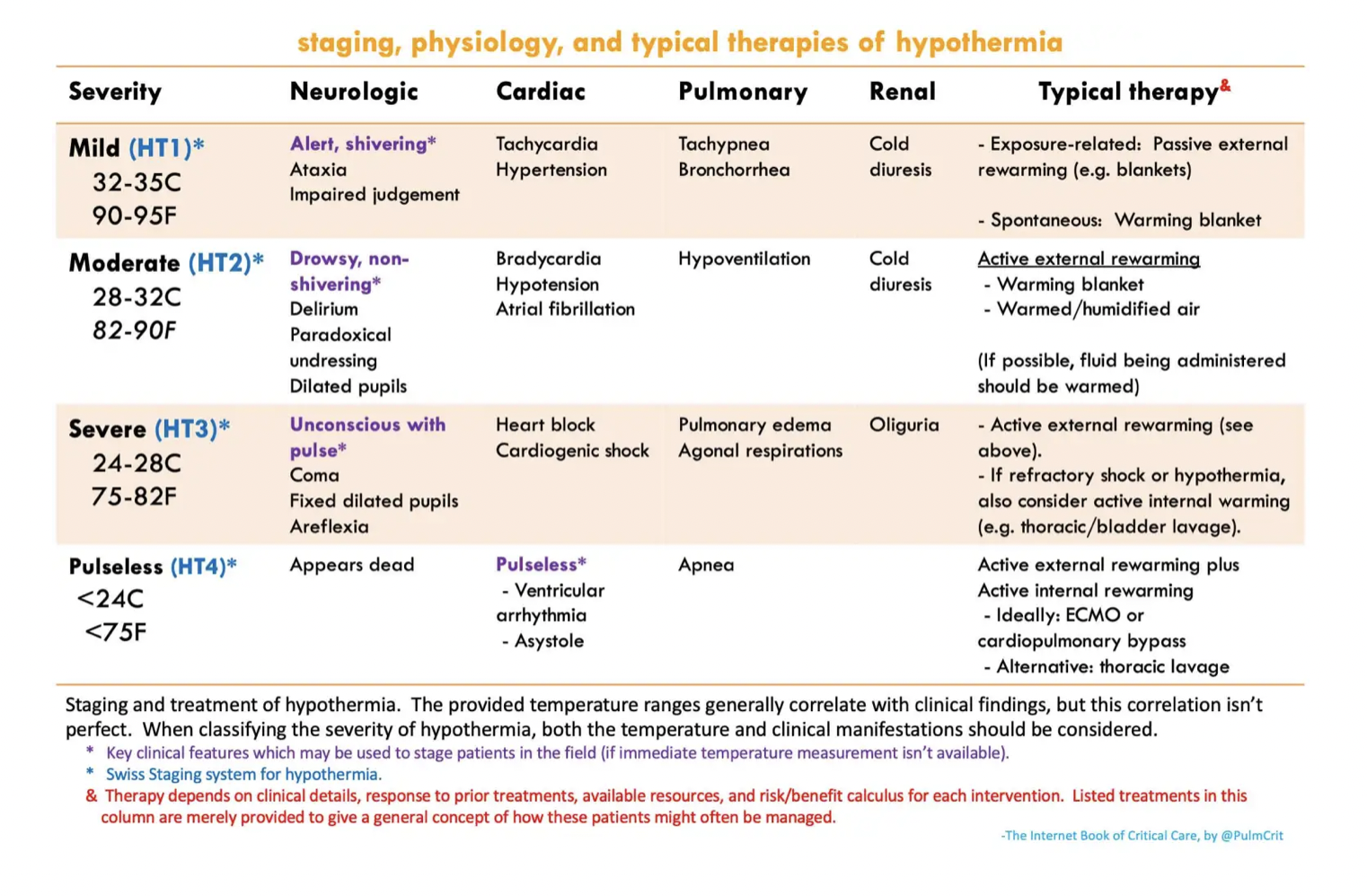
Swiss Hypothermia Staging System:

Stage 1: Mild (32-35°C) - Shivering, mild confusion, awake

Stage 2: Moderate (28-32°C) - Severe shivering, altered mental status

Stage 3: Severe (20-28°C) - Loss of consciousness, bradycardia, shivering may cease

Stage 4: Profound (<20°C) – Unobtainable vital signs

Associated Complications:

  1. Cardiac dysfunction

    1. Dysrhythmias can occur when body temperature drops below 30°C

    2. There is typically a drop in temperature and MAP after rewarming is started due to vasoconstriction

  2. Cold injuries (frostbite, etc. Maybe there will be more on this at a later date)

  3. Coagulopathy (patient may be coagulopathic despite normal labs because the lab rewarms the sample)

    1. Impaired clotting function

    2. Thromboembolism (due to hemoconcentration and poor circulation)

    3. DIC

  4. Impaired pharmacology

    1. Protein binding increases when temperature drops, rendering drugs ineffective

    2. Oral meds are not absorbed well due to decreased GI motility

    3. IM route is impaired due to vasoconstriction

  5. Rhabdomyolysis

General Management:

  1. Airway, Breathing, Circulation (ABCs)

    • Hypothermia causes a leftward shift in oxygen curve so support with oxygen, and prepare for intubation depending on how profound the hypothermia is

  2. ECG Findings

    • Patients usually have sinus bradycardia, can progress to a fib with slow ventricular response

    • Severe cases can develop v fib

    • Osborn or "J" waves (associated with moderate to severe hypothermia)

  3. Remove Wet Clothing - Prevent further heat loss

  4. Passive External Rewarming - Insulate the patient, provide warm blankets

  5. Active External Rewarming (should be done for moderate hypothermia)

    • Use forced warm air blankets or radiant heaters – our ED uses the Bair Hugger

  6. Active Internal Rewarming (for severe hypothermia)

    • Warmed intravenous fluids (warmed to 38-42°C)

    • Heated humidified oxygen

    • Various lavages (Thoracic, peritoneal, bladder, GI)

Management during Cardiac Arrest:

  1. CPR – initiate if patient does not have a pulse (should also assess if patient is still breathing)

    • It is challenging to assess vital signs in hypothermic patients - use end tidal or POCUS to help assist to see if patient is breathing and has cardiac function

    • Starting CPR if the patient does have a pulse may precipitate ventricular rhythms

    • Hypothermic patients have higher chances of improved neurological outcome and survival than normothermic patients that arrest

  2. Defibrillation

    • Use defibrillation if indicated, but note that hypothermic patients may not respond to defibrillation until adequately warmed

  3. ECMO

    • Patients with refractory hypothermia should be considered for ECMO

    • Patients with out-of-hospital-cardiac-arrest that are hypothermic should ideally be transported to an ECMO center

    • If patient is unstable (dysrhythmia, severe hypothermia, etc) ECMO teams should be contacted early in the ED visit


Stay warm out there this weekend!


Paal P, Pasquier M, Darocha T, Lechner R, Kosinski S, Wallner B, Zafren K, Brugger H. Accidental Hypothermia: 2021 Update. Int J Environ Res Public Health. 2022 Jan 3;19(1):501. doi: 10.3390/ijerph19010501. PMID: 35010760; PMCID: PMC8744717.

Baumgartner EA, Belson M, Rubin C, Patel M. Hypothermia and other cold-related morbidity emergency department visits: United States, 1995-2004. Wilderness Environ Med 2008;19:233-237

Brown et al., Accidental Hypothermia. N Engl J Med 2012; 367:1930-1938


POTD: Primary Hypothermia!

With the turn of the weather and puffy jacket season in full throttle, let's review hypothermia. In this POTD, we're going to be focusing on primary (environmental) hypothermia, and not hypothermia due to secondary causes such as sepsis, hypothyroidism, metabolic derangements, or trauma. 


Hypothermia is defined as core temperature < 35C


  • between 1991-2011, an average of 1,300 deaths/year in the US attributed to environmental hypothermia

  • At risk populations: elderly, lack of shelter, alcohol/drug abusers, frequent exposure (winter sports)


The modified staging system for hypothermia classifies severity into 4 categories, of which the chart below summarizes and demonstrates the general clinical picture


Initial evaluation should include the following:

  • Basic vital signs including core temperature

  • BGM (hypoglycemia)

  • EKG (bradycardia, Osborne waves, arrhythmias)

  • Basic labs (hypokalemia)

  • lactate, CK (rhabdomyolysis)

  • coags and fibrinogen (DIC)

  • consider TSH, cortisol, 


Management of ABCs:

Airway/Breathing

  • Intubate as necessary, however encourage holding off intubation as patients are often hypotensive and prone to arrhythmias. Attempt rewarming and stabilization prior to hemodynamic stressors of intubation.

Circulation

  • bradycardia 

    • hypothermia itself causes bradycardia, thus the key to treating bradycardia is rewarming. Giving medications to speed up HR is generally not encouraged and can induced arrhythmias

  • hypotension 

    • again, often hypothermia induced, may also be secondary to bradycardia. Rewarming should improve pressures, and vasopressors generally discouraged as it can also induced arrhythmias

    • BUT - vasodilation can occur with rewarming, so if there is a drop in pressure, vasopressors should be considered

  • access 

    • if central access is needed, keep the guidewire shallow to prevent entry into the right ventricle. The hypothermic heart is incredibly sensitive to VT/VF, so don't go tickling that ventricle. Femoral access or midline access is preferred


EKG in hypothermia:

Patient may initially present with tachycardia, however as hypothermia sets in, everything gets slower. Just imagine taking a NSR rhythm stripe and click-dragging it horizontally. You often see bradycardia, prolonged QTC, prolonged PR, and the presence of Osborne waves. 

  • Osborne waves (also known as J waves) - a deflection at the junction of the QRS and ST segments, present usually temp < 30C. The size increases with worsening hypothermia

The most common dysrhythmia is atrial fibrillation, but cardiac arrest is due to eventual VF/VT or asystole

Management of hypothermia in the pulseless patient:

The famous phrase: "They aren't dead unless they're warm and dead" indeed is true. To be considered dead, the patient's core temperature should be ~32C before calling it.

ACLS: Modifications in setting of hypothermia

  • Delayed or intermittent CPR can be adequate due to low oxygen demand. Some say for T < 28, there can be pauses of <5 minutes CPR after every 5 minute interval of CPR if necessary. 

  • Medications may fail to metabolize and will accumulate in the system, therefore prolonged intervals between epinephrine pushes and limiting repeated doses are recommended. 

  • Similarly, defibrillation is poorly effective in T < 30C and repeated defibrillation may induce myocardial damage. 

    • Suggested treatment by AHA: Can attempt one defibrillation if VF/VT present, otherwise hold further defibrillations and all IV medications until core T > 30C

Internal Rewarming:

  • ECMO: The best option for sever hypothermia. It allows for better organ perfusion, active rewarming as much as 7-10C per hour, and allows you to stop compressions. As an ECMO center, we should definitely consider getting ECMO downstairs to cannulate.

  • Thoracic Lavage: achieved through placement of two chest tubes, left preferred over right if only able to place one tube. Can achieve 3-6C warming/hour. Use Belmont or warm tap water if available, and monitor appropriate ins and outs to prevent tension PTX from improperly draining tubes

  • Bladder Lavage: less effective, however is easier to implement. Use dedicated 3-way Foley catheter or consider instilling 300 cc warmed fluids, hold for 15 minutes, and draining bladder. Rinse and repeat.

Management of hypothermia in patient with pulse:


  • External rewarming: Removing cold/frozen clothes and fully wipe down of snow. Employ external rewarming with Arctic Sun (preferred due to direct contact with skin to improve rewarming), warm blankets or Bair Hugger. 

  • Respiratory rewarming: Warmth is typically lost through expirations. Therefore consider rewarming via respiratory support. In non-intubated patients, HFNC or CPAP/BIPAP with highest temperature settings. Intubated patients should have heated and humidified air set up with the ventilator (ask respiratory)

  • Fluid rewarming: hypothermia induced "cold diuresis," therefore patients often require volume resuscitation. Warm IV fluids, ideally crystalloid, should be administered. Keep in mind that this method prevents further dropping temperatures, but is ineffective for raising core temperature. 

  • Lavage can also be considered in moderate/severe hypothermia in patients with pulses - see above.


Resources:

https://emcrit.org/ibcc/hypothermia/

http://www.emdocs.net/em3am-hypothermia-2/

https://www.saem.org/about-saem/academies-interest-groups-affiliates2/cdem/for-students/online-education/m4-curriculum/group-m4-environmental/hypothermia

https://wikem.org/wiki/Accidental_hypothermia


POTD: Winter is Coming.

Let’s talk about FROSTBITE, BRRRRRRR.

Background

  • Results from the freezing of tissue that are exposed to temperatures below their freezing point, resulting in direct ice crystal formation and cellular lysis with microvascular occlusion

  • Most of the damage occurs as a result of a freeze thaw cycle with endothelial damage and cellular death resulting in osmotic gradient changes, initiation of the arachidonic acid cascade, vasoconstriction, and hematologic abnormalities including thrombosis

  • Risk correlated with temperature and wind speed

  • Risk is <5% when ambient temperature (includes wind chill) is > –15°C (5°F)

  • Most often occurs at ambient temperature < –20°C (–4°F)

  • Wetness and humidity increase the risk (water has 25x thermal conductivity of air)

  • Can develop within 2-3sec when metal surfaces that are at or below –15°C (5°F) are touched

  • Most commonly affects distal part of extremities, face, nose, and ears

  • The severity of irreversible damage is most closely related to ambient temperature and length of time the tissue remains frozen

  • High-risk groups: outdoor workers, elderly, homeless, drug or alcohol abusers, psychiatric disease, high-altitude or cold-weather athletes, military personnel

  • "Hunter's response" - prolonged repeated exposure to cold is protective

Classification

Frostbite is classically categorized into four levels of injury.

Screen Shot 2021-10-18 at 1.04.25 PM.png

Management

The initial treatment in the Emergency Department for all degrees of frostbite is the same. Addressing ABC’s, trauma evaluation, removing wet and constrictive clothing, treatment of concomitant hypothermia (must rewarm to a core temperature of at least 35°C), and identification of other injuries should be confirmed in all cold injury cases if warranted.

  • THAWING: Do NOT attempt until the risk of refreezing is eliminated. Refreezing will cause even more severe damage. Rapid active rewarming is the core of therapy and should be initiated as soon as possible. Best performed in a circulating water bath around 37°C to 39°C. Frostbitten faces can be thawed using warm water compresses, and ears may be thawed with small bowls of warm water. Immersion rewarming can be discontinued when the affected area developed a red or purple appearance and becomes pliable to the touch.

  • Analgesia: rewarming is very painful, treat your patient's pain!

  • Local wound care: Gently dry, elevate, and apply bulky dressing to the affected area. Compartment syndrome is a known complication, so maintain a high suspicion.

  • Update tetanus as needed

  • Empiric prophylactic antibiotics are not needed and are controversial.

  • Surgical management may be required if wet gangrene or infection occurs, but this is typically reserved for late frostbite management after the rewarming phase in days to weeks following initial presentation

Dispo Dispo Dispo

  • Patients with superficial local frostbite may be discharged home if social circumstances allow. Patients unable to care for themselves adequately should never be discharged into subfreezing temperatures.

  • Significant injuries will require admission.

References:

http://www.emdocs.net/brrr-ed-presentation-evaluation-and-management-of-cold-related-injuries/

http://emedicine.medscape.com/article/926249-treatment#showall

https://wikem.org/wiki/Frostbite

https://www.emrap.org/episode/environmentalem/hypothermia

https://www.emrap.org/episode/environmentalem/frostbite