Approach to a pt with thrombocytopenia

Definition: definition of thrombocytopenia is a platelet count < 150 × 10E9/L


Presentation:

Often asymptomatic when first detected (significant or spontaneous bleeding rarely occurs with a platelet count > 50 × 10E9/L; 5-fold increase in risk <50 x 10E9/L)

Purpura, petechiae and bleeding from the gums and into the knee

Bleeding from venipuncture sites

Spontaneous intracerebral haemorrhage (<0.5% in ICU, higher risk if platelets <10 x 10E9/L)

Splenomegaly


Petechiae

Petechiae


Purpura

Purpura


Etiologies


Increased destruction

Immunologic: collagen vascular disease, infection, ITP, lymphoma/CLL, drugs (heparin, sulfonamides, aspirin, phenytoin, digoxin, vancomycin, B-lactam antibiotics)

Mechanical: TTP, HUS, DIC, chronic dialysis patient

Vasculitis


Decreased production

Decreased megakaryocytes: drugs (EtOH, thiazide, chemo, linezolid), toxins, infection, leukemia

Splenic sequestration


Dilutional

Massive transfusion, ECMO, exchange transfusion


Suggested work-up:

Repeat platelet count – Ensure value is accurate

CBC – TTP-HUS presents with anemia AND thrombocytopenia. Abnormal platelets and abnormal WBC count is concerning for primary hematologic etiology.

Peripheral smear (call the lab and ask them to add a peripheral smear) – Look for schistocytes, and also for platelet clumping to rule out pseudothrombocytopenia. 

PT/PTT/INR/fibrinogen – These will be abnormal in DIC and unaffected in other etiologies.

Consult Heme/onc


Whom to consider admitting:

Severe thrombocytopenia (i.e. < 20)

Platelet count drop > 50% of baseline

Suspected leukemia, TTP-HUS, or DIC

Active bleeding


Diagnosis to consider:

HITT

Heparin-induced thrombocytopenia AND thrombosis

Use the 4T score: Timing (5-10 days after heparin exposure), degree of Thrombocytopenia (> 50% drop from baseline), no other clear etiology, and Thrombosis

If intermediate probability, send anti-platelet (PF4) antibody test

If high probability, start alternative anti-coagulation with argatroban (hepatically-cleared) or bivalirudin (renally-cleared)

ITP

Dx of exclusion

Acute => child, viral prodrome days-weeks prior, platelets <20, self-limited, supportive care unless active bleeding (steroids, IVIG, anti-Rh Ig)

Chronic => adult, no prodrome, gradual, platelets 30-100

Life-threatening bleeding: plt transfusions, steroids, IVIG


TTP-HUS

Usually idiopathic but can be 2/2 meds (clopidogrel, ticlopidine, quinine) or infection (E. coli)

Thrombocytopenia + microangiopathic hemolytic anemia (pentad of fever, anemia, thrombocytopenia, renal involvement, and neuro involvement)

Acute management is with plasmapheresis

If not available FFP and arrange for transfer. NO PLATELETS.


Leukemia

Look for high white counts, but sometimes can present with leukopenia

Patients should be started on all-trans retinoid acid (ATRA) as soon as the diagnosis is suspected

When to transfuse platelets: <10 or <50 + active bleeding or <50 + invasive procedure. Of note: transfused platelets last 3-5 days.


 · 
Share

Pulmonary hypertension

Pulmonary Hypertension (PH): 

pulmonary hypertension news.jpg

Pulmonary hypertension (PH) is present when mean pulmonary artery pressure exceeds 25 mm Hg at rest or 30 mm Hg with exercise (Normal PA systolic pressures range from 10-30)

Definitive diagnosis via right heart catheterization, rare cause of SOB

In general, there is no cure besides supportive care and treating the precipitant, high rate of mortality


Pathophysiology:

Pulmonary vasculature is meant to be a high-flow, low-resistance circuit.

Cardiac causes

LA or LV disease => ↑ LA pressure => ↑ pulmonary venous pressure => ↑ pulmonary artery pressure => ↑pulmonary vascular resistance

L to right shunt will also cause high pulmonary vascular pressure


Respiratory causes

hypoxic vasoconstriction -> PH


Which leads to => vasoconstriction => altered vascular endothelium and smooth muscle function

 => cellular remodelling => increased vascular contractility => lack of relaxation in response to various endogenous vasodilators => fibrosis of vascular tissue


Symptoms: 

Dyspnea (with rest or with exertion), Fatigue, Chest Pain, Syncope, Exertional lightheadedness

Patients with severe pulmonary HTN can develop signs of R heart failure (JVD, hepatomegaly, ascites, edema)


Five types of PH: 

Group 1: Pulmonary arterial HTN

Idiopathic

Genetic/Heritable abnormalities

Drug/Toxin induced

Associated with known risk factors (HIV, liver disease, collagen vascular disorders)


Group 2: Pulmonary venous HTN (left heart disease)

Systolic or diastolic dysfunction

Mitral or aortic valve disease


Group 3: Chronic hypoxemic lung disease

Obstructive lung disorders (COPD)

Interstitial Lung Disease

Idiopathic Pulmonary Fibrosis

Sleep-Disordered breathing (OSA)


Group 4: Embolic disease (PE)

Group 5: Miscellaneous


The Workup

EKG:

Most common abnormality is right axis deviation

Signs of R heart strain: S1Q3T3, right atrial enlargement in the inferior leads, incomplete/complete RBBB

Labs:

CBC, CMP often nonspecific

BNP often elevated and correlates with outcomes

Elevations in troponin are associated with higher morbidity and mortality

CXR

Can demonstrate signs of RV failure – enlarged RA, RV, pulmonary arteries

Can demonstrate underlying etiology – hyperinflation, ILD, edema

Echo

Best initial diagnostic test

Apical four chamber helpful to evaluate size of RV relative to LV and assess for septal deviation

US not helpful in assessing volume status in these patients!


Management

Start with ABC’s 

Give home medications

If known PH consult pulmonology early


Consider 3 Ps: Preload, Pump and Pipes

RV function determined by 3Ps: Preload, Pump, Pipes

Preload:

Consider gentle hydration (250cc IVF) vs. gentle diuresis 

Pump:

Cardiovert dysrhythmias as indicated

Consider inotropic support

Dobutamine 2-10 mcg/kg/min

Milrinone 50mcg/kg bolus -> 0.2-0.8mcg/kg/min (can cause hypotension)

Consider low dose norepinephrine (0.05–0.75mcg/kg/min) to maintain coronary artery perfusion

Pipes: (afterload)

Consider Pulmonary vasodilators

Prostanoids, endothelin receptor antagonists, and phosphodiesterase-5 (PDE-5) inhibitors

Prostanoids are treatment of choice

Epoprostenol is the only therapy proven to improve survival

inhaled nitric oxide 20-40ppm (good in bypass, doesn’t cause systemic hypotension as inactivated when bound to Hb)

Treat underlying etiologies


If need to provide respiratory support consider doing it in consultation with pt’s pulmonologist:

Always start with NRB before positive pressure ventilation (PPV)

PPV can cause rapid cardiovascular collapse due to increased Pulmonary Vascular Resistance and decreased preload

If you must intubate, have vasopressors at bedside: phenylepherine + NE + vasopressin are all OK

Low TV, low PEEP ventilation strategy

aggressively treat hypercarbia, acidosis, hypothermia (all increase PVR), which can increase pulmonary vascular resistance, pulmonary artery pressure, and RV strain


References: LITFL, EMDocs, UpToDate


 · 
Share

systematic approach to reading CXR and hidden pneumonias


For this pearl of the day we will talk about systematic approach to reading CXR and hidden pneumonias:


The key is to be very systematic when approaching CXRs and that is what radiologists do each time.

Here is the suggested approach by the Brown EM program (https://brownemblog.com/?offset=1533674064239&category=Education)

Screen Shot 2019-08-16 at 2.07.54 PM.png


https://commons.wikimedia.org/wiki/File:Mediastinal_structures_on_chest_X-ray.svg#/media/File:Mediastinal_structures_on_chest_X-ray,_annotated.jpg

When ready to review the x-ray, consider the commonly used “A, B, C, D, E, F” system.

A - Airway- trachea, carina, right and left main bronchi

B - Bones and soft tissue- clavicles, ribs- posterior and anterior, vertebral bodies, and sternum on lateral films. Look for any fractures, dislocations, or lytic lesions.

C - Cardiac- cardiac silhouette and mediastinum. The cardiac silhouette should be less than half of the thoracic cavity. AP films exaggerate heart size, so this rule does not apply. Assess the borders of the heart and the hilar structures

D - Diaphragm- right should be higher than left and you should see a gastric air bubble on the left. Is there any free air under the diaphragm? Evaluate the costophrenic angle and pleura (normally invisible due to thinness).

E - Everything else (lines and tubes, pacemakers, artificial valves)

F - Fields- FINALLY, evaluate the lung fields. Lungs are the area of greatest interest, so it is helpful to keep this at the end to prevent distraction. Divide each lung into three “zones” when reading a chest x-ray. These do not correlate with the lobes. Remember, there are 2 lobes on the left (upper and lower) and 3 on the right (upper, middle and lower). 


Hidden pneumonias:

Go through your ABCDEFs and look at the signs of hidden pneumonias:


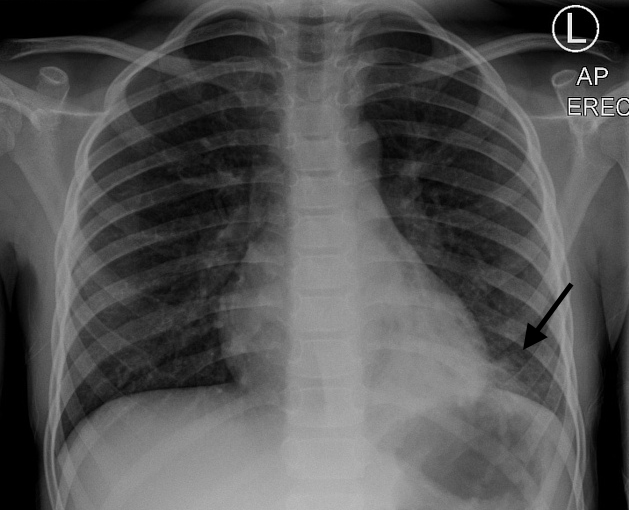
Silhouette sign

The loss of the normal silhouette of a structure is called the silhouette sign.  - It enables us to find subtle pathology and to locate it within the chest.

Screen Shot 2019-08-16 at 2.09.18 PM.png


Screen Shot 2019-08-15 at 4.16.26 PM.png

R middle lobe pneumonia

2a.jpeg

LLL pneumonia

Screen Shot 2019-08-15 at 4.27.05 PM.png

LLL pneumonia


Hidden areas

There are some areas that need special attention, because pathology in these areas can easily be overlooked:

apical zones

hilar zones

retrocardial zone

zone below the dome of diaphragm

These areas are also known as the hidden areas.


But in doubt get another view or a chest CT.


References:

https://brownemblog.com/?offset=1533674064239&category=Education

http://www.radiologyassistant.nl/en/p497b2a265d96d/chest-x-ray-basic-interpretation.html#in5145a34e91e18

https://www.bir.org.uk/media/258608/mark_rodriguez_-_philips_trainee_for_excellence_-_unofficial_guide_to_radiology.pdf