Alternative Headache Therapies

Hi everyone! For today's POTD, let's think outside the box when it comes to acute headache management in the ED. Headaches in general are one of the most common chief complaints we see in the ED. We have our tried-and-true drug cocktails -- an antidopaminergic drug (ex: reglan, phenergan, compazine), +/- benadryl, +/- toradol -- but what happens if those fail?

1) Other drug therapies: a) Sumatriptan 6 mg subcutaneously, or 10-20 mg intranasally [exercise caution in pts with cardiovascular disease] b) Dihydroergotamine 0.5-1 mg IM or IV [nausea is common; also exercise caution in pts with cardiovascular risk factors] c) Valproic acid: 300-1200 mg IV

2) Sphenopalatine ganglion block: Decent amount of anecdotal support for its efficacy (although no large studies yet), and very easy to do! - Soak long cotton-tipped applicators in your choice of local anesthetic - Insert nasally and apply firm pressure until you meet resistance at the posterior wall of the nasopharynx - Leave in place for 5-10 min; bask in the glory of your migraine-stopping powers https://www.aliem.com/2017/03/trick-sphenopalatine-ganglion-block-primary-headaches/

3) Occipital nerve block: https://www.youtube.com/watch?v=JGLOaZpZwqU - Consider this for patients with primarily occipital distribution of their pain! - 1 mL of 0.5% bupivacaine injected over the greater and/or lesser occipital nerves (located along the imaginary line between the occipital protuberance and the mastoid process, as shown in this photo)

4) Cervical paraspinous nerve block: https://www.youtube.com/watch?v=oy1lggvxV9Y - Similar concept to the occipital nerve block; consider this for your cervicogenic headache patients - 1.5 mL of 0.5% bupivacaine injected approx 2-3 cm bilaterally to C6-C7, to a depth of 1-1.5 inches

References: https://www.aliem.com/2016/05/managing-migraine-headaches-complicated-patients/ http://www.emdocs.net/headache-management-best-current-evidence-ed/ http://rebelem.com/alternative-headache-therapies/ http://www.annemergmed.com/article/S0196-0644(16)30301-8/pdf https://www.ncbi.nlm.nih.gov/pmc/articles/PMC3737484/
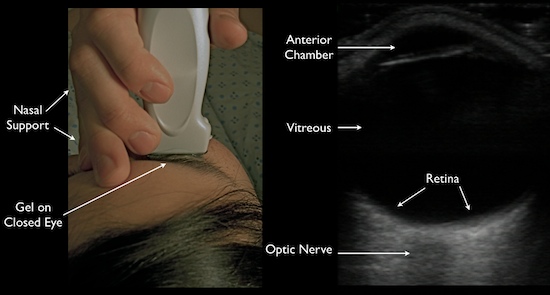
 · 
Share

Ottawa Aggressive Protocol

For today's POTD, we're dusting off our analytical skills and looking at an important paper from 2010 that attempted to settle the age-old debate of how to best manage symptomatic acute-onset (<48 hr) atrial fibrillation, using a strategy known as the Ottawa Aggressive Protocol. The actual paper is available here (https://www.cambridge.org/core/services/aop-cambridge-core/content/view/S1481803500012227), but here is a bite-sized summary for you all, because 'tis the season of giving.

First things first: What is the Ottawa Aggressive Protocol (OAP)? The OAP study aimed to show that rapid cardioversion followed by discharge home was a safe and effective option for those ED patients presenting with clear acute onset a-fib (defined below as <48 hr duration) who were clinically stable.

Study population: 660 adult patients presenting to a single Ottawa hospital with symptomatic atrial fibrillation for <48 hr

Exclusions: Patients with chronic AF, symptoms > 48 hours or of unknown duration, and/or patients with another primary diagnosis requiring hospital admission.

Intervention: All 660 patients got an initial attempt at pharmacologic conversion with IV procainamide (1 g infused over 1 hour). If the patient had a history of failure to respond to procainamide, an alternative IV antiarrhythmic agent could be used before moving on to electrical cardioversion in the ED. Patients with a history of multiple unsuccessful prior attempts at pharmacologic cardioversion using all available drugs proceeded directly to electrical cardioversion. Attempts at rate control (with IV metoprolol or diltiazem) were performed in those patients who were either highly symptomatic or not planning to undergo cardioversion.

Design: Retrospective, consecutive cohort study

Key results: 58.3% of patients successfully converted with procainamide. 91.7% of patients successfully converted with electrical cardioversion. 96.8% of patients were discharged home; of those 93.3% were in normal sinus rhythm. At the 7-day mark, the relapse rate was only 8.6% (i.e. patient found to be back in a-fib). There were zero cases of thromboembolic events, torsades, or death. Median ED length of stay was 4.9 hours (3.9 hr for those pts who converted after procainamide; 6.5 hr for those requiring electrical cardioversion).

Weaknesses: Not a RCT; no blinding; only 7 days of follow up.

What does it mean for my practice? The Ottawa Aggressive Protocol is a reasonable approach for management of AF of <48 hours duration, and may help decrease lengths of stay and resource utilization (sparing a hospitalization). Patients were safely discharged after cardioversion (procainamide +/- electricity) without need for systemic anticoagulation or antiarrhythmic prophylaxis, with arrangements for outpatient cardiology follow-up.

References

Stiell et al. Association of the Ottawa Aggressive Protocol with rapid discharge of emergency department patients with recent-onset atrial fibrillation or flutter. CJEM, 2010 May;12(3):181-91.

CoreEM.net

WikEM.org


 · 
Share

Ocular Ultrasound

For today's POTD, we'll be taking a closer l👀k at eye pathology, specifically how we can use ocular ultrasound to further evaluate many common eye complaints encountered in the ED.

While eye complaints are fairly common in the ED, our comfort level with the diagnostic tools available to us for these complaints varies greatly.
These tools include:
- Slit lamp (the hardest part is really finding and flipping the 4 separate switches to actually turn it on)
- Fluorescein and Wood's lamp
- Tonopen (addressed in a previous POTD)
- Bedside fundoscopy (I'll admit I suck at this)
- And, ocular ultrasound!
The biggest advantage of ocular sono is how easy it is to perform.
1) Slap on a tegaderm over the patient's closed eye
2) Squeeze on a generous amount of ultrasound jelly
3) Gently lay on your linear probe and look both horizontally and vertically (tell the patient to look straight ahead behind their closed eyes)

You can make so many diagnoses using ocular ultrasound!!!
- Foreign body
- Globe rupture
- Lens dislocation
- Retinal detachment
- Vitreous detachment
- Increased ICP (seen through increased optic nerve sheath diameter: measure 3 mm down, then 5 mm across)

We'll quickly focus in on retinal vs vitreous detachments. This is explained wonderfully in video form at http://5minsono.com/rdvd/, which I will summarize here.


In a retinal detachment, you can see the lines connectingto the optic nerve*. [How to remember this: the retina is just an extension of the optic nerve]

*To get a little more technical, you can also use ultrasound to tell whether a retinal detachment is "mac-on"(macula still attached, i.e. need for emergent surgical reattachment) or "mac-off" (macula detached, i.e. damage probably already done).
- The key to understanding this is knowing that the macula is located lateral to the optic nerve (or in eye-speak, on the temporal side).
- This 1 minute video illustrates the difference better than I can in words: https://www.youtube.com/watch?v=JijIfSzOG9U
- I think in reality though, if I suspect any kind of retinal detachment in a patient, whether it's mac-on or mac-off, I'll be transferring them for emergent ophtho evaluation.


vitreous detachment is typically seen floating above and NOT connected to the optic nerve. It is often also hazier and swirlier (very technical terms), especially as the patient looks in different directions.

References:
Academic Life in EM
5MinSono.com
EMin5.com
 · 
Share