Superficial Cervical Plexus Block POD

That’s right. Time to talk about my favorite nerve block.

The SCPB

This block is 

quick

and

easy

.

It makes

IJ central lines

painless procedures

.

It also provides excellent analgesia for

clavicle fractures

,

ear lobe lacerations

,

blind subclavian lines

, or

anything within this quadrilateral

:

boundries.jpg

How to do it

Find the

posterior aspect of the sternocleidomastoid (SCM) muscle

.

Position the probe half way down the SCM as you measure it from mastoid process to clavicle.

About at the level of the superior aspect of the thyroid cartilage, also about where the EJ crosses over the sternocleidomastoid.

land-marks-jpeg.jpg
with-probe-jpeg.png

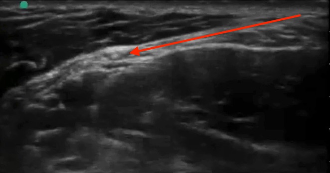
The fascial plane under the posterior aspect of the sternocleidomastoid muscle is your target.

just-sono-arrow.jpg

Like other

plane blocks

 you are not targeting any one nerve in particular. By infiltrating this tissue plane, you get the superficial plexus as it peeks out from behind the SCM at this level:

PastedGraphic-2.tiff

Inject 5-10 cc of local anesthetic.

Ensure it is spreading in the plane like this.

1.jpg
2.jpg
3.jpg

Safety:

  • As with all ultrasound guided nerve blocks, visualize your needle tip always, especially prior to injection.

  • When you begin injecting, inject one mL only to ensure you see it spreading in the fascial plane. Then inject the rest.

  • Withdraw before you inject if there is even a slight possibility you are close to a vessel

  • Throw some color on your site to ensure you identify any vessels.

  • In general this is a very well tolerated and forgiving block.

  • The incidence of phrenic nerve involvement is extremely low with SCPB, far lower than with the interscalene block. Phrenic nerve involvement will be avoided if ensure you don’t go too deep - a few cm, or about half way down the deep edge of the SCM. Going deeper than this results in a deep cervical plexus block which will result in some motor and sensory blockade of the arm.

  • Horner’s syndrome is a rare and self-limiting complication.

  • The amount of local anesthetic used in this block is nowhere near close to toxic levels, so local anesthetic systemic toxicity will not occur as long as you manage to avoid the IJ and carotid.

Tips and troubleshooting

  • For a central line, you can set up, gown, drape, and set up your ultrasound like usual, then use the 5 cc lidocaine which come in the central line kit for your block. Place the block first, then flush your line/lay out your equipment, and your patient will likely be completely numb by the time you’re ready to start your line placement.

  • The other option is to place your block while you’re doing your pre-scan. This way you can use 5-10 cc, but you have to get it out of the Pyxis. Just clean the probe and the skin with a chlorhexidine swab and use sterile gel.

  • If it’s your first time doing a block, consider doing it as a 2 person block. Use some IV extender tubing and have another provider operate the syringe for you. I like slightly longer tubing than typical IV tubing, e.g. the one below. At Maimo, you can find it in the stock room between North and South sides, top shelf straight ahead when you first walk in.

  • Can't visualize your needle? Make sure your ultrasound probe is directly above it and in-line with it. Next, make sure it is as close to parallel to the surface of the probe/perpendicular to the ultrasound beams as possible. If you are approaching a 45 degree angle, your needle will be close to invisible. At close to a 0 degree angle, it will shine like a laser beam.

  • You can use the 27 gauge needles to make it more comfortable but they are a little harder to see.

Further reading and references:

http://highlandultrasound.com/superficial-cervical-plexus-block/

 - Highland crushes nerve block education with their website and their SCPB page is no exception

https://www.ultrasoundpodcast.com/2015/03/superficial-cervical-plexus-block-with-bedsidesono-trust-us-this-is-really-awesome-foamed/

 - Mike and Matt of ultrasound podcast also did an amazing episode on this where many of the images in this tutorial are from

https://www.nysora.com/cervical-plexus-block

 -

Another good resource

 · 
Share

Crashing Asthmatic POD

We treat asthma on a daily basis, especially in the peds ED. But what if duonebs x3, steroids, and mag isn’t doing the trick?

THE CRASHING ASTHMATIC

  • Nebulized epinephrine may help.

  • If no improvement, start dosing IM epi as if it were anaphylaxis: 0.5 mg (0.01 mg/kg) q 10 min, or start a drip at 5 mcg/min and titrate to effect.

  • Keep going with continuous albuterol nebs.

  • If pharmacy isn’t around to make an epi drip, consider a “dirty” epi drip: 1 mg epi (an entire vial of code cart epi) added to 1 L NS or LR, start at 2 drops per second and titrate up.

  • Alternatively, terbutaline IV can be started: 10 mcg/kg bolus over 10 min and then drip starting at 0.4 mcg/kg/min and titrate up. Terbutaline is a systemic beta agonist. Perhaps they’re so tight that the albuterol you’re nebulizing is not getting where it needs to go due to profound bronchoconstriction. The main adverse effect is here is vasodilation-related hypotension.

By this point, your intubation stuff should be ready and the patient should be in resus.

They also will be having insensible losses so should get as 20 cc/kg IVF bolus.

Still getting worse.

Now this gets interesting.

We are really trying to avoid intubating any asthmatic because of historically poor outcomes with intubation, but sometimes it is unavoidable.

Next step is to try BiPAP. BiPAP could also be started simultaneously with epi. If they can’t tolerate BiPAP, consider ketamine to help them tolerate BiPAP. Ketamine can be dosed numerous ways. If sub-dissociative dosing is pursued, you risk them freaking out. If dissociative dosing, there’s a higher risk of laryngospasm. But consider this, they’re on the brink of getting intubated anyway. If your last-ditch-effort-ketamine gives them laryngospasm, that might be your cue to push a paralytic.

Ketamine and BiPAP has failed.

Time to intubate. Preoxygenate as much as possible. Use the largest ETT possible. First pass success is key. Induce with ketamine 2 mg/kg if they’re not already in the K-Hole. Roc or Sux.

Now they’re intubated

  • They have OBSTRUCTIVE LUNG PHYSIOLOGY. It will take them way longer than usual to exhale. Thus:

  • Low respiratory rate! 8 breaths/min

  • Lung protective tidal volume: 7 cc/kg ideal body weight

  • Minimal PEEP: 0 (ZEEP) - 2 cc H2O

  • High inspiratory flow rate: 90 LPM or I:E 1:5

  • FiO2 100%

  • The ventilator will alarm due to high PEAK pressures. This is OK. Have the respiratory therapist fix it to raise the alarm threshold. The high peak pressures are a consequence of their tight bronchioles.

  • If running into issues with ventilator dyssynchrony, consider paralyzing with cisatracuium

  • Relative hypoxia (sat mid 80s, goal >90%) and hypercarbia (goal >7.15) is OK

  • Aggressive airway suctioning

  • If they begin crashing, disconnect from vent and push on chest to ensure breath stacking is not the issue; rule out pneumothorax; rule out displaced/clogged/kinked ETT

Still doing poorly

  • Call the ECMO team for VV ECMO

  • Anesthesia to set up inhaled anesthetics! e.g. desflurane, sevoflurane. Not tons of evidence, but in case series' and anecdotally, this works really well.

  • Fun fact, CO2 can be dialyzed out of someone rather than ventilated out of someone. However, you need to be in a center where ECMO is also done, because it’s basically putting a piece of the ECMO circuit into a CVVHD circuit. Yes. Blew my mind too.

See Reuben’s algorithm on this at https://emupdates.com/when-the-patient-cant-breathe-and-you-cant-think-the-emergency-departement-life-threatening-asthma-flowsheet/

 · 
Share

Nephrotic Syndrome Pearl of the Day

Nephrotic Syndrome

Pathophysiology

Think about it like this: Glomerular basement membrane problem, you spill all your protein (albumin) from your plasma into your urine, leading to high urine protein, low serum protein, and edema. It may help to simply think of it as decreased oncotic pressure causing movement of fluid from the intravascular space to the interstitial space; in reality the pathophysiology of edema in nephrotic syndrome is a little more complicated - has to do with a combination of primary (due to renal disease) and secondary (via RAS pathway) sodium retention.

Thus,

nephrotic syndrome is defined by:

1. Heavy proteinuria

(protein excretion > 3.5 g/24 hours; UPEE>3.5; >2 in children)

2. Hypoalbuminemia

(< 3 g/dL; <2.5 in children)

3. Peripheral edema

(Hyperlipidemia and thrombotic disease are frequently observed with nephrotic syndrome but not required for the diagnosis, may also be immunosuppressed)

(UA should have no significant hematuria, casts, or RBCs which would suggest a nephritic picture)

Measuring Proteinuria 

The most convenient way is to calculate a 

Urinary Protein Excretion Estimation (UPEE)

(insert infantile bathroom humor here)

UPEE (g/day) = (urine protein (mg/dL)) / (urine creatinine (mg/dL))

This works because a random urine protein to urine creatinine ratio very closely approximates the true 24 hour urine protein excretion, as shown below.

Image result for urine protein to creatinine ratio

Interpretation:

UPEE <2.0 g/day —> Within normal limits

UPEE 2.0–3.5 g/day —> Above normal limit - investigate further

UPEE >3.5 g/day —>Nephrotic range

Nephrotic vs Nephritic

Remember to always think of nephrotic syndrome in contrast to nephritic syndrome! I love this image:

Image result for nephrotic nephritic

Workup

1.

Make the diagnosis: urinalysis, urine protein, urine creatinine, serum albumin, a lipid panel, basic metabolic panel

2. Consider further testing to differentiate primary vs secondary on a case-by-case basis: HIV, ANA, complement (C3/C4 and total hemolytic complement), serum free light chains and urine protein electrophoresis and immunofixation, syphilis serology, hepatitis B and hepatitis C serologies, and the measurement of cryoglobulins; when in doubt, run it by nephrology

3. Consider testing for complications: POCUS for pleural effusion/ascites; CXR for pleural effusion, dopplers or CTA for venous thromboembolism; antithrombin III, plasminogen, protein S (hyper coagulability); immunoglobulins; 

3. Usually renal biopsy is required for definitive diagnosis

Etiology/Treatment/Dispo

In children 10 years or younger, it is minimal change disease (MCD) 90% of the time. Most MCD responds to corticosteroids.

In children >10 years, it is MCD >50% of the time.

In adults focal segmental glomerulosclerosis (FSGS) is the most common etiology (35%). It can be primary/idiopathic, or associated with other disease processes, most commonly HIV or massive obesity.

Untitled.jpg

In most cases, a biopsy will be needed in order to confirm diagnosis.

Admit patients with severe edema, pulmonary effusions or respiratory symptoms, or signs and symptoms suggestive of systemic infection or thrombotic complications to the hospital.

Discharge with nephrology follow up ASAP and low salt diet if only mild-moderate edema.

In kids age 1-10, may consider starting a course of steroids (

Prednisone 2 mg/kg/day x 6 wks then 1.5 mg/kg every other day x 6 wks) only if:

  • Age 1-10

  • No renal insuficiency

  • No macroscopic hematuria

  • No sx systemic disease

  • No HTN

  • Normal C3 levels

Special scenarios

Nephrotic syndrome + chest pain?

DDX: PE and myocardial ischemia because hyperthrombotic, pneumonia (immunosuppressed), pleural effusion

Consider POCUS, CXR, CTA, EKG, cardiac enzymes

Severe edema:

Requires lasix

May need albumin infusion prior to lasix if anasarca or signs of intravascular depletion

Hopefully you’ve already consulted ICU if you’re having to do this

Significant hypertension:

ACEi or ARB

SBP/Empyema:

Pleural effusions/ascites are common in severe fluid overload

Both are extra susceptible to infection in this state

Low threshold for diagnostic paracentesis/thoracentesis

References

Uptodate: Overview of heavy proteinuria and the nephrotic syndrome

Tintinalli’s Emergency Medicine: A Comprehensive Study Guide, 8e: Chapter 134: Renal Emergencies in Children

Michael Mojica, MD. “PEM Guides.” NYU Langone Medical Center, 2015. iBooks. 

https://itunes.apple.com/us/book/pem-guides/id1039923332?mt=11

Google Image Search

 · 
Share