VOTW: Can’t shrug this one off!

Hello all! This week’s VOTW is brought to you by myself!

Hospital course

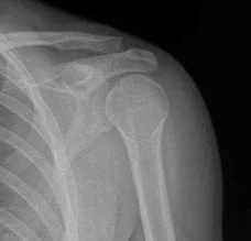
An 18 y/o M presented after falling off his skateboard causing him to land on his left shoulder. The XR showed a posterior shoulder dislocation, seen in the XR below where we can see the classic light bulb appearance of the humeral head.

Ultrasound

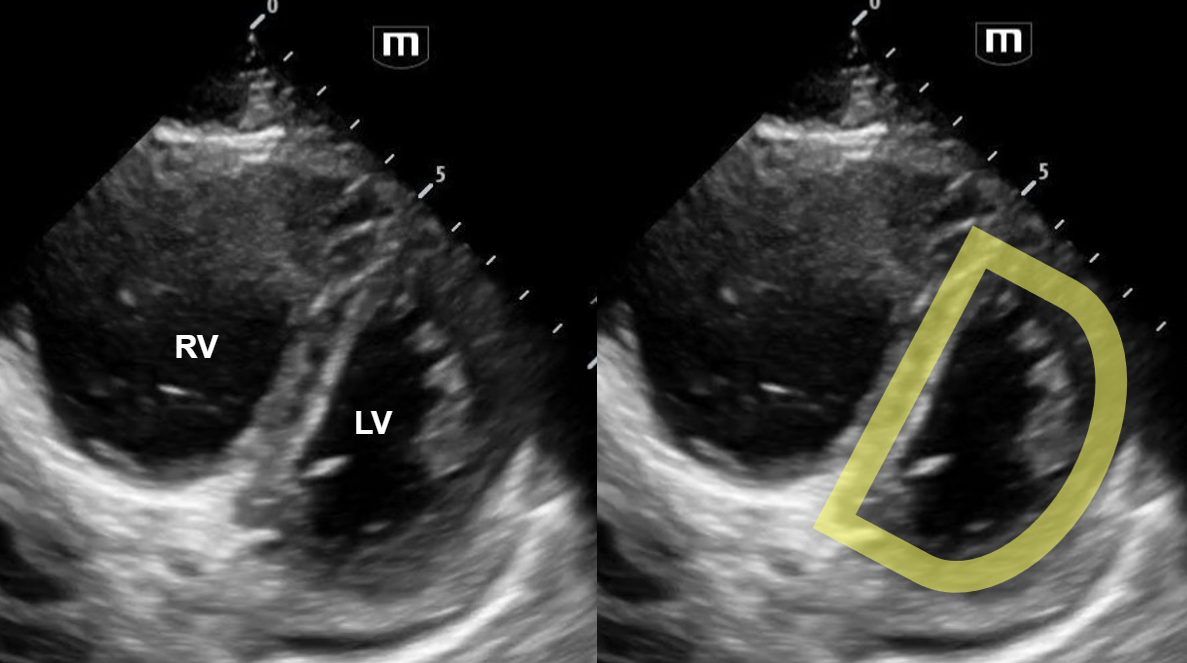
The left shoulder was scanned with ultrasound, which is shown below.

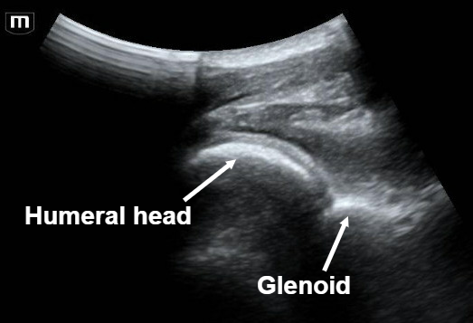
Note that close proximity of the humeral head to the probe (more superficial), which is indicative of a posterior shoulder dislocation. Also note that the glenoid fossa is not seen well, indicating that the humeral head is not articulating well with the glenoid.

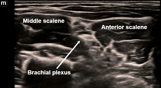
An US-guided interscalene nerve block was completed to relieve the patient’s pain. The target is the brachial plexus cords seen in the image above between the middle and anterior scalene muscles.

In the above clip, we can see the needle entering in-plane lateral to medial with injection of local anesthetic within the interscalene space, surrounding the brachial plexus.

Case Conclusion

After the interscalene block, the patient had great pain control and the shoulder was able to be reduced successfully without any procedural sedation!

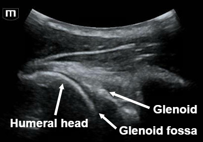
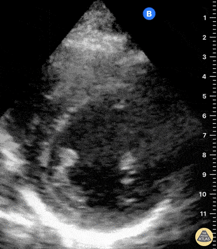
This ultrasound of the shoulder was obtained post-reduction. Here we can see that the humeral head is articulating directly with the glenoid.

In this clip, we can see the glenohumeral joint with the humeral head moving well in normal alignment as the shoulder adducts and abducts.


Posterior shoulder dislocation

  • A posterior shoulder ultrasound scan is done by placing the probe just over the scapular spine in a transverse plane, with the probe marker towards the patient’s left.

  • A posterior shoulder dislocation will show the humeral head displaced closer to the probe (appears more superficial on the screen). Also, the humeral head will not be articulated with the glenoid fossa.

  • An US-guided interscalene block can be performed in the ED as a replacement for procedural sedation prior to shoulder reduction as seen with this patient! The interscalene block covers the shoulder and proximal humerus. The probe is placed 2-3 cm superior to the clavicle and the target is the brachial plexus which is commonly referred to as the “stoplight” sign in this view because of its 3 circular hypoechoic structures found between the middle and anterior scalene muscles.


Happy scanning!

Sono team

Resources to review:

 · 

TASER

Happy Tuesday! I hope you all enjoyed your Labor Day Weekend. 

For this week’s trauma Tuesday topic, I wanted to discuss Tasers. In the past 2 years I have seen a handful of patients in NYPD custody in need of medical checks as a result of being tased. 

Taser guns were first developed in the 1970’s. The name is actually an acronym which stands for Tom A. Swift Electric Rifle. The inventor, Jack Cover, was a huge fan of the science fiction book series featuring the main character Tom Swift. The Taser was introduced for use by law enforcement in the 1990s. It falls under the category of less-lethal weapons which also includes batons, mace, tear gas, and projectiles like foam rounds.

The Taser is a type of Conducted Electrical Weapon. Pulling the trigger releases the compressed nitrogen. The 2 pronged darts that are attached to a copper wire fire out. As the darts make contact with skin, the electrical circuit is completed and electricity courses through. As it does, it causes painful involuntary muscle contraction. 

As of 2019, the American Academy of Emergency Medicine does not recommend routine workup in an otherwise well appearing and asymptomatic individual who was exposed to less than 15 seconds of electrical current. No need for EKG’s, laboratory testing or observation. There has been little evidence showing significant medical harm or physiologic change in patients who have received a Taser shock, even in those who have underlying cardiac abnormalities or defibrillators.

It is important to assess the skin and area that was struck by the prongs. Those can cause minor burns and skin injury. There have been isolated case reports of the metal barbs causing penetration injuries as well. This is a case study where a patient had a barb stuck in his penis. Also consider other injuries that someone may sustain if they had gotten tased and fallen onto the ground. 

References

  1. https://www.aaem.org/statements/evaluations-after-a-taser-device-activation/ 

  2. https://www.britannica.com/topic/TASER 

  3. Baliatsas C, Gerbecks J, Dückers MLA, Yzermans CJ. Human Health Risks of Conducted Electrical Weapon Exposure: A Systematic Review. JAMA Netw Open. 2021 Feb 1;4(2):e2037209. doi: 10.1001/jamanetworkopen.2020.37209. PMID: 33576818; PMCID: PMC7881359. 

  4. Theisen K, Slater R, Hale N. Taser-Related Testicular Trauma. Urology. 2016 Feb;88:e5. doi: 10.1016/j.urology.2015.11.011. Epub 2015 Dec 1. PMID: 26592466. 

  5. Bralow LM. Misfire: A man with genital trauma. J Am Coll Emerg Physicians Open. 2022 Dec 21;3(6):e12875. doi: 10.1002/emp2.12875. PMID: 36570370; PMCID: PMC9772490.

 · 

VOTW: “D”-oh! What a heart!

Hello all! This week’s VOTW is brought to you by myself.

Hospital course

30 y/o M presents to the ED after 2 syncopal episodes. He had 10 days of worsening dyspnea on exertion with chest pressure and palpitations. He flew to California 1 month ago and returned yesterday.  

In the parasternal short view of the heart above, we see two cardiac chambers, the right ventricle (RV) and the left ventricle (LV). We can see flattening of the interventricular septum towards the LV chamber, creating a “D”-shaped LV (labelled above).

View the attached clip to see the LV take on a shape of the letter “D” with each contraction! Also note that the RV is dilated and appears larger in size than the ”D”-shaped LV.

The clip above shows a parasternal short view of a normal heart. Notice that the left ventricle appears circular, and the right ventricle forms a smaller crescent-shape surrounding the left ventricle.

Case Conclusion

CT imaging showed pulmonary emboli within the bilateral pulmonary arteries and dilatation of the right atrium and right ventricle associated with right heart strain.

Thrombectomy was deferred because patient was hemodynamically stable. He was started on a heparin infusion in the ED and then admitted.   

Right heart strain

·       Pulmonary embolism can cause an acute increase in pulmonary pressures and right ventricular afterload that causes increased right heart strain. Focused ultrasound has been shown to be both highly sensitive and highly specific in detecting anatomical changes of the heart seen with right heart strain.

·       “D-sign” is a finding suggestive of right heart strain seen on the parasternal view of the heart. Increased right ventricular pressures cause bowing of the interventricular septum into the LV, causing the “D”-shaped LV to form.

·       Other findings suggestive of right heart strain include increased RV size, McConnell’s sign, and TAPSE.


Happy scanning!

Sono team

PS: just in case nobody gets it, the subject line pun is a Simpsons reference!


Resources to review:

·       https://www.thepocusatlas.com/right-ventricular-dysfunction/

·       https://everydayultrasound.com/blog/category/Right+Ventricular+Strain

·       https://www.acep.org/sonoguide/basic/cardiac

 ·