Unstable Pelvic Ring Fractures

The pelvic ring consists of the sacrum and two innominate bones, which are made up of the pubis, ilium, and ischium. These bones are held together by strong ligaments to give the pelvis stability.

A pelvic ring fracture is a severe fracture with 2 breaks in the circular ring, leading to an unstable pelvis and a potentially unstable patient. Fractures that disrupt the pelvic ring predispose patients to bleeding given the large network of arterial and venous anastomoses. Patients who have an isolated pelvic fracture and are hypotensive carry a mortality of 15-40%. Most vascular injuries in the pelvis are venous (90%). While rare, arterial bleeds (10%) should be suspected when a pelvic binder is placed but the patient remains hemodynamically unstable. The retroperitoneal space can accumulate 4 liters of blood before venous tamponade occurs. Pelvic binders are useful in that they can help tamponade bleeding veins, decrease total pelvic volume, and prevent the shifting of bony fragments.

Other unstable pelvic fractures include lateral compression fractures, "open book" pelvic fractures, and vertical shear fractures. Lateral compression fractures occur when a lateral force vector (t-bone in an MVC) causes an anterior ring disruption and sacral fracture.

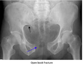
“Open book” fractures occur as a result of anteroposterior compression injury to the pelvis, commonly caused by high-speed trauma or elderly falls. There is a disruption to the pubic symphysis and the pelvis opens like a book. Diastasis of > 1 cm (blue arrow) can indicate instability. Disruption of the pubic symphysis, one of the strongest ligamentous structures in the human body, requires a lot of force and should be a red flag to look for other injuries to the head, spine, chest, or abdomen.

Vertical shear pelvic fractures are seen when one-half of the pelvis shifts upward as a result of a fracture of ipsilateral anterior and posterior pelvic ring fractures. They typically occur as a result of high-energy force applied in the axial direction (aka from the gas pedal to the femur and up to the pelvis). Patients may have an unstable pelvis and leg length discrepancy.

For all unstable fractures, you should appropriately resuscitate and stabilize the patient. Give blood as needed but avoid transfusing through lower limb access because it may drain into the retroperitoneal space. If there is a pelvic ring fracture, consider binding the pelvis. Your binder should lay over the greater trochanters and have enough force to close the pelvic ring (video:https://www.youtube.com/watch?v=tWLBZKeWEkg).


EMS Protocol of the Week: Eye Emergencies

Hey all,

This week's protocol looks at eye emergencies that apply to both the adult and pediatric populations.

The prehospital approach starts with CFRs at the most basic level to evaluate and initiate treatments based on these ocular findings:

1) Non-penetrating foreign objects/chemical eye injuries: flush affected eye with NS for 20 minutes

2) Impaled object to eye: use bulky dressings to stabilize object and cover eye to prevent consensual eye movements

3) Avulsed eye: cover eye with saline, sterile dressings and do NOT place eye back into socket

BLS providers provide the additional support of removing contact lenses as needed.

ALS providers provide the additional support of administering proparacaine 0.5% or tetracaine 0.5% drops for chemical eye injuries to assist with irrigation.

Not alot to do on the OLMC side other than to help assist our EMS providers in each ocular scenario.

Check out www.nycremsco.org or the protocol binder on North Side for more.

John Su

PGY-2


POTD: Le Fort Fractures

Hello everyone!

For trauma Tuesday, let's discuss Le Fort fractures.

Le Fort fractures are complex facial fractures involving the maxilla, zygoma, and orbital rims. They were discovered by Dr. Rene Le Fort who discovered these "lines of weakness" in skulls of patients with blunt facial traumas. These fractures by nature include the pterygoid structures of the sphenoid bone, which provide stability and support for the mid face. Most commonly seen in MVC, the velocity determines the severity of the fractures, of which there are three categories:

Le Fort I: "floating palate"

- a transverse fracture of the maxillae above the teeth, leaving the body of the maxilla separated from the pterygoid plate and nasal septum. This leads to a "floating palate", where the maxilla and hard palate may be mobile.

- associated with malocclusion and dental fractures

- generally considered a stable fracture

Le Fort II: "floating maxilla"

- fracture that extends superiorly to include the nasal bridge, maxilla, and orbital rim and floors. fractures are typically bilateral and appear triangular in shape

- The maxilla and nose are mobile, the eyes/orbits are not

- can be stable or unstable

Le Fort III: "floating face"

- the rarest and most severe, this fracture involves the bridge of the nose, medial and lateral orbital wall, zygomatic arch, and maxilla. 

- the entire face is mobile, can present as a "dish face" deformity (essentially the face is caved in)

- this is an unstable fracture

Presentation and Evaluation:

Le Fort fractures can present with many features, including facial deformity and emphysema, CSF rhinorrhea, conjunctival hemorrhage, raccoon eyes, hemotympanum and auricular hematoma, and anosmia

Questions to ask:

Can you smell? Can you bite? 

How is your vision?

Is there numbness or tingling in you face?

Exam:

- palpate for signs of crepitus, areas of tenderness, or instability

- visual acuity test - very important considering high risk of ophthalmologic damage

- check mobility by stabilizing the forehead and grabbing the upper teeth/hard palate, and attempt to move the hard palate

-evaluate to c-spine injuries - approximately 1.4% have concomitant c-spine injuries or dislocations

Management:

- Stabilize ABCs. If airway is at risk - understand that it will be a difficult airway, and consider awake intubation. These patients are particularly difficult as oral injury may prevent appropriate jaw displacement for oral intubation. Nasal intubations are contraindicated due to nasal injuries. These are patients where if a definitive airway is needed, cricothyroidotomy should be considered.

- significant nasal bleeding can occur and may present an airway risk. Consider anterior packing and elevation of head of bed to 40-60 degrees. Posterior packing should be avoided due to risk of skull base injuries.

- IV antibiotics should be given in sinus fractures or CSF leaks, which will be the majority of these fractures

- CT with dedicated facial view should be obtained. 

Disposition:

- All Le Fort fractures should be seen by OMFS

- consider Ophtho or NSG consult if there is concern for eye or brain damage/CSF leak

- some stable Le Fort I and II are stable for discharge with follow up, however most will require ICU (for airway management) or direct OR 

http://www.emdocs.net/em3am-le-fort-fractures/

https://www.ncbi.nlm.nih.gov/books/NBK526060/

https://coreem.net/core/le-fort-fractures/