And a Happy New Year of Wellness

Today's pearl will be brief and focus on Wellness. We are often super busy on our shifts and in our lives, but it doesn't take much time to stop - pause - and add a little wellness to your day. 

Here are 9 ways to have less stress in under a minute.

  1. Breathe. Take a big breath in and feel your stomach muscles relax. Hold it for a moment. Purse your lips, and blow your breath out slowly as if you are blowing out candles on a birthday cake. Feel your stomach muscles tighten as you empty your breath. Do it again 2 times or more if needed.

  2. Smile- even if you don’t feel like smiling! Smiling has been shown to decreases stress response, and even shown to lower heart rates during a stressful situation. So maybe every so often a little  "grin and bear it" can help reduce your stress!

  3. Relax your Mouth. Open your mouth, let your jaw and tongue relax, or try a jaw massage. Often times stress will lead to jaw clenching. Relaxing your jaw helps signal to your brain to reduce your stress response. 

  4. Laugh! Laughing decreased your stress levels and long-term may even boost your ability to fight sickness.

  5. Give others or yourself a hug. Hugging help reduce stress for both the giver and the receiver!

  6. Shrug your shoulders. Bring your shoulders up to your ears for a slow count of 5, then release.

  7. Exercise. Do some quick squats, jumping jacks, running in place, walk across the street to grab some tea or water, etc.

  8. Peel an orange – seriously! Then inhale the smell. Research shows that the smell of citrus relaxes people.

  9. Say Thank You. Really. Think of things you are grateful for. It is another great stress reducer! 

Wishing you a safe, happy, and healthy New Year!

References:

https://www.smithsonianmag.com/science-nature/simply-smiling-can-actually-reduce-stress-10461286/

Simply Smiling Can Actually Reduce Stress | Science | Smithsonianwww.smithsonianmag.comA new study indicates that the mere act of smiling can help us deal with stressful situations more easily
https://exploreim.ucla.edu/wellness/stressed-it-may-take-a-toll-on-your-teeth-explore-an-integrative-approach-to-managing-bruxism/
https://www.mayoclinic.org/healthy-lifestyle/stress-management/in-depth/stress-relief/art-20044456
https://health.usnews.com/health-news/health-wellness/articles/2016-02-03/the-health-benefits-of-hugging
https://nihrecord.nih.gov/newsletters/2006/02_24_2006/story03.htm

https://www.ncbi.nlm.nih.gov/pubmed/22071630
https://nycwell.cityofnewyork.us/en/coping-wellness-tips/less-stress-in-under-a-minute/
https://www.psychologytoday.com/us/blog/what-mentally-strong-people-dont-do/201504/7-scientifically-proven-benefits-gratitude

 · 

Do you see what eye see?

Do you see what eye see?

Eye complaints can be abundant in the ED - so what should we focus on? Today's pearl is going to focus on the diagnoses of atraumatic vision loss.

Let's break it down by symptoms: Painless or painful?

Good physical exam must include: visual acuity, visual fields, pupillary exam, fluorescein woods lamp, slit lamp and of course - a head to toe complete physical exam. 


Painless Vision Loss: 

  1. TIA/stroke: sudden vision loss usually from embolic or thrombotic event, can often be from carotid artery occlusion. If transient, often called amaurosis fugax but that can apply to any cause of transient vision loss. Risk factors: it is a stroke/TIA of the eye, so same cardiovascular risk factors. Testing: MRI, ECG, Echo, Carotid US. ED role: Don't miss it - this is a stroke and should admitted to a monitored stroke unit.

2. Central retinal artery occlusion: Sudden, permanent typically unilateral vision loss (permanent damage ~90 minutes of occlusion). Risk factors: atherosclerosisdisease (HTN, DM, Smoking, etc.) and treatment involves treating risk factors (varied success with laser treatment of pressure decreasing eye drops); or secondary to Giant cell arteritis (+/- headache, pain with hair burshing or chewing)  in which case rapid high dose corticosteroids can prevent vision loss. PE: decreased visual acuity, asymmetric red reflex, "Cherry red spot" on the macula on fundoscopy. ED role: urgent/emergent optho referral or ED consult. Recognize if cause is giant cell arteritis (CRP, ESR) and treat with high dose steroids. Consider timolol 0.5% topical drops but no strong evidence. Will need PCP to address concurrent risk factors. 

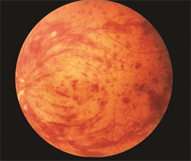
3. Central retinal vein occlusion: often times sudden but occasional gradual (days-weeks) vision loss due to swelling of the macula. Risk factors: same as above and glaucoma. Think of this as a DVT for the eye. PE: decreased visual acuity. "Blood and thunder" on fundoscopy. ED role: Evaluate for cause or presence of other pathologies secondary to a hypercoagulable state (DVT, PE, sickle cell, etc.). Refer to optho and will need PCP to address concurrent risk factors. 

retinal vein occlusion image-full.png

4. Retinal Detachment: Sudden, spontaneous "flasher and floaters" or "spots or stars" or loss of vision that is like a "curtain closing." Usually loss of peripheral vision. Risk factors: recent eye injury or surgery, severe nearsightedness (think elderly). ED Role: POCUS - look for a delicate floating line in the anechoic eyeball space. If their is no macular separation, their vision can be saved! This is an optho emergency. Unless you're Errel, err on the side of caution and call optho for all of these since we might not be able to definitively determine if the macula is involved.

5. Vitreous Hemorrhage and Posterior Vitreous Detachment: Floaters, strings, or cobwebs in their vision that change with eye movement. Flashes of light. Risk factors: retinal damage (surgery, trauma, prior retinal tear), Diabetic Retinopathy, trauma, sickle. ED role: POCUS: swirling cloud-like opacity at moves with ocular movement and is not tethered to the optic disk or retina "washing machine sign". ED role: Avoid Anticoagulation, elevate head of bed, optho referral.

6. Optic Neuritis/papillitis: painless vision loss over hours to days, typically unilateral. +/- prior episodes. +/- other neuro symptoms. May worsen with eye movement. Risk factors: typically females 18-45, multiple sclerosis. Viral: mono, zoster, encephalitis, TB. Physical exam: decreased acuity, relative afferent pupil defect, often normal fundoscopy; requires complete neuro exam. ED role: MRI brain. Diagnose underlying cause. Often required admission for continued IV high dose steroids. 

Painful Vision Loss:

1. Acute angle glaucoma: sudden, usually form bright to dark environment (movie theater). Often with headache, N/V, and light sensitivity/halos. PE: midsize, nonreactive pupil. Must include tonometry, IOP>20! Risk factors: Asian, femaile, shallow anterior chamber. ED role: timolol  0.05% 1-2 drops (beta blocker, watch for systemic absorption SE) eye drops and brimonidine (alpha agonist) eye drops to reduce IOP. Miotic agent pilocarpine (2-4% 1-2 drops q15 m). Titrate until symptoms improve, IOP decreases. Systemic carbonic anhydrase inhibitors like acetazolamide 500 mg IV. Elevate head of bed. Treat nausea and headache, too! Urgent optho referral.

2. Corneal Ulcer: foreign body sensation, red eye. PE: injected conjunctiva, gray patch on cornea. Increased fluorescein uptake on Woods lamp. Possible hypopyon purulent collection. ED role: Evaluate for foreign body. Consider HSV keratitis. Start opthalmic antibiotic drops, must have pseudomonas coverage for contact lens wearers (tobramycin), consider antifungal. **Admit for IV ceftriaxone for suspected gonococcal infection. Will need optho evaluation and ulcer culture. 

cornealulcer.jpg
Corneal-Abrasion.jpg

3. Uveitis/Iritis: painful, progressing red eye worse with eye movement, +photophobia suggests anterior. Floaters suggests posterior, often no pain. Can be panuveitis. Causes can be inflammatory (HLA B-27 usually bilateral, eg. reactive arthritic, psoriasis, IBD, ankylosing spondylitis; sarcoid, Kawasaki), traumatic, infectious (toxoplasmosis, CMV, HSV, adeno, measles, mumps, TB, syphilis, Lyme), or secondary to medications(sulfa). PE: injected conjunctiva with ciliary flush (erythema around the iris). Sluggish, constricted, or irregular pupils. Slit lamp shows cloudy anterior chamber and "cell and flare." ED role: evaluate for underlying cause (STD screen, CXR for TB, etc.) and refer to optho urgently and appropriate f/u. 

I am certain that this is not a comprehensive list, so please - learn on!

Buzzword pearls to part with: 

OpthalmologySession_web.jpg
 · 

Holy smokes! Inhalation Injury

First steps (stable pt)
-treat as a trauma pt (ABCDE) and look for traumatic injuries
-place pt on NRB with O2 to 15 L/m
—add nebs 4% lidocaine early to prepare for visualization of cords with videolargynoscope
4:2:1 rule for burn pt fluid resuscitation
—give fluids even if no external burns visible, as pt will have insensible losses
-treat pain!
-evaluate cords and surrounding laryngeal structures for edema with video laryngoscope or bronch

How to risk-stratify your patient with suspected smoke inhalation injury:

Screen Shot 2018-12-27 at 12.04.46 PM.png

Rule out
carbon monoxide toxicity: obtain serial blood gases (send co-oximetry) to monitor carboxyHb. Normal levels are 5 – 12%, depending on whether or not the pt is a smoker. Also consider if family presents with similar vague symptoms.
-cyanide toxicity: cyanide levels are not reliable in excluding toxicity, as it is rapidly cleared, and don’t result for days. Use lactate>8 or rising lactate despite fluid resuscitation to raise suspicion for toxicity
—ddx for elevated lactate (=impaired tissue oxygenation) in burn pt: cyanide, metHb, hypoxia, volume depletion
-look for rhabdo and AKI

Warning signs of respiratory failure
drooling or difficulty swallowing = impending failure
-monitor for stridor, hoarseness, and respiratory distress
-PaO2/FiO2 ratio indicates degree of pulmonary shunting past injured lung. PaO2/FiO2<300 forewarns respiratory failure

Intubation
-early elective intubation in a controlled setting is better than crash intubation of a pt with edematous airway structures
-Prepare multiple sizes ETTs in anticipation of vocal cord edema. Use the largest that will fit so that the pt can get a bronchoscopy upstairs. Prepare suction for soot-filled secretions. Sux is safe to use up to 24h post-burn.
-use volume controlled ARDS settings (6-8 mL/kg TV)
—airways and lung become less compliant in inhalational injury, so must prevent barotrauma and allow for permissive hypercapnea

 ·