Local Anesthetic Systemic Toxicity (LAST)

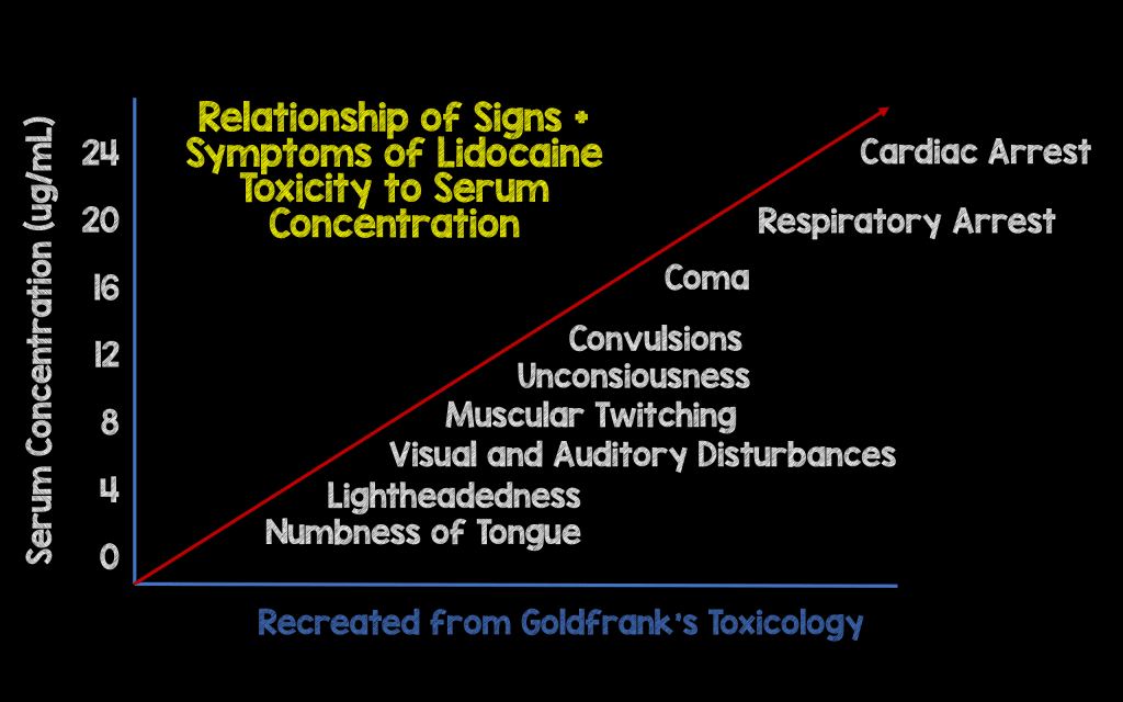
So a guy comes in with 16 superficial stab wounds overnight and you get to be the lucky resident to sew him up (true story). Halfway through, he starts complaining his mouth is numb (this part didn't happen). What do you do? LAST can occur from three important etiologies - direct systemic injection, accumulation from multiple injections, or injection into a highly vascularized area. Most importantly, prevent this from happening by knowing your max dose, aspirating before injecting, and asking about symptoms.

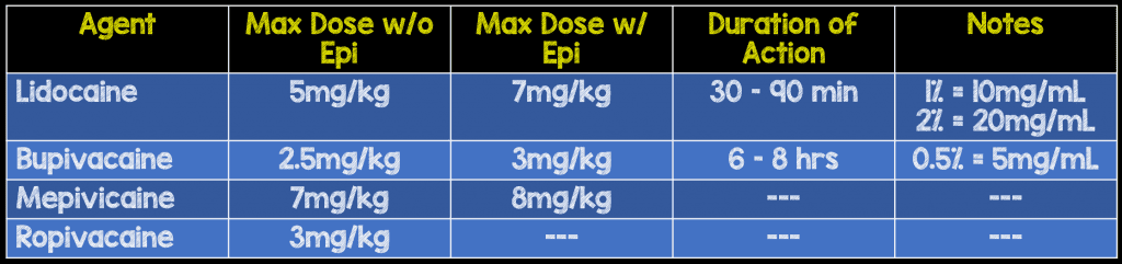
Remember local anesthetics are sodium channel blockers, so when introduced systemically cause cardiac and neuro toxicity.


First, stop injecting the patient. Then,

A - You may need to intubate. B - Give 100% FiO2 and hyperventilate as acidosis, hypoxemia can push this syndrome into cardiac arrest. C - Hypotension should be treated with IVF and push-dose pressors (epi). In arrest, high-quality CPR should be continued for a while as these anesthetics are lipid soluble and take time to redistribute. Consider pacing and ECMO. D - Seizures can occur and benzodiazepines are the treatment, as usual.

If the patient has an arrhythmia, hemodynamic instability, prolonged seizure, or rapid progression of symptoms, break out the 20% intralipid. Call pharmacy/look up the dose - 1 ml/kg over 1 minute q3m x3 and then 0.25 ml/kg/min. Theoretically, lipid emulsion therapy works by drawing anesthetic out of the plasma.

Image result for lipid sink


Sources

Rebel EM: http://rebelem.com/local-anesthetic-systemic-toxicity-last/

LITFL: Local Anesthetic Toxicity

Wiki EM: Local Anesthetic Systemic Toxicity

Kapitanyan, R. Local Anesthetic Toxicity. Medscape http://emedicine.medscape.com/article/1844551-treatment#d1

 · 

Hypothermia EKG

Hi all, winter is coming,

Image result for jon snow dying
The above patient is brought into Resus 52 by EMS as a notification for both a stroke and STEMI. You aren't able to a history as he appears obtunded, and you obtain the following EKG.
An Osborn wave (late delta wave, J-wave) is a characteristic (but not pathognomonic) finding of hypothermia at temperatures lower than 32C. It is a positive deflection at the J-point (between QRS complex and ST segment) most prominent in the precordial leads. There is correlation with the degree of hypothermia and the magnitude of the osborn wave which resolves with rewarming. Other situations this can occur are hypercalcemia, neurological injury, certain medications, and as a normal finding.  Other EKG findings in hypothermia are shivering artifact, bradycardia, and prolonged PR, QRS, ST segments.
Typical Osborn waves
Mild hypothermia (32-35C) presents as shivering and some drowsiness. In these patients, initiate passive external rewarming measures (warm blankets).
Moderate hypothermia (28-32C) presents as loss of shivering and progressive lethargy. Vitals signs begin to be affected with a drop in HR and cardiac output.
Severe hypothermia (<28C) presents with coma and severely depressed cardiopulmonary function. Arrhythmias and cardiac arrest (a-fib, v-fib, asystole) start to occur at this point as well. Rewarming may be needed to achieve ROSC. For moderate and severe hypothermia, you'll want to use active external rewarming (Bair Hugger) and active internal rewarming. For the latter, methods include warm IV fluids, warmed humidified air, bladder lavage, gastric lavage. On the more extreme side you could also do peritoneal lavage, pleural lavage, or ECMO (most rapid rewarming technique).
(subtle resolution of J wave)
Sources
LIFTL, Osborn Wave; lifeinthefastlane.com/ecg-library/basics/osborn-wave-j-wave/
Smith, Steve Osborn waves and hypothermia. hqmeded-ecg.blogspot.com/2011/11/osborn-waves-and-hypothermia.html
Rosh Review

 · 

Unstable Cervical Fractures

Today's POTD is on trauma, which will be the topic every Tuesday. The mnemonic to remember unstable C-spine fractures and dislocations is "Jefferson bit off a hangman's thumb."

Jefferson burst fracture - Axial loading force (diving) transmitted to C1 causing a bilateral fracture of the ring.

- Associated with other cervical fractures, vertebral aa injury.


Bilateral facet dislocation - Hyperflexion injury (rapid deceleration) causing anterior dislocation of superior vertebral body by 50% of the body's AP diameter. - Anterior and posterior ligaments are disrupted.


Odontoid type II and type III fractures

- Fracture of the dens of C2 - Type II - fracture through waist, or base of odontoid near attachment to C2 - Type III - extension of fracture to the upper portion of body

Image result for type 3 odontoid fracture

(type 3)


Atlanto-occipital dissociation

- Called "internal decapitation" because it is frequently fatal.

- Flexion injury with injury to ligaments stabilizing atlanto-occipital joint.


Hangman's fracture - Extreme hyperextension injury - MVC, diving, judicial hangings. - Bilateral C2 pedicle fracture, causing anterior dislocation of C2 vertebral body onto C3.


Teardrop fracture - Hyperextension of the anterior longitudinal ligament avulsing a teardrop fragment of the body.

- Or hyperflexion causing the vertebrae bodies to collide and form a teardrop fragment of the superior vertebrae displacing the body and disrupting the posterior longitudinal ligament.


Sources

Rosen's (ed 8), ch 43

Rosh Review

Young, N, et al. Unstable spine fractures (wikem.org/wiki/Unstable_spine_fractures)

 ·