Supracondylar Fractures

sc9.png

Pediatric Supracondylar Fractures

Background

  • Defined by fracture of distal aspect of humerus above the epicondyles

  • Mechanism

    • Direct: blow to the elbow, fall onto flexed elbow

    • Indirect (more common): FOOSH, fall onto hyperextended UE

      1. 95% of these fractures are due to extension injury

  • Most common age: 5-8 year olds

    • Also more likely to be dislocated in this age group

  • Males>Females

Exam

  • Complain of pain/swelling/decreased ROM of elbow

  • “S shaped deformity”

    • when fracture is entirely displaced (distal humerus)

  • Need to perform neurovascular exam!

    • Median nerve: A-OK sign

      1. Mostly commonly affected

    • Radial nerve: thumbs up sign

    • Ulnar nerve: abduct/adduct fingers (try to remove paper they are holding in between adducted fingers)

  • Check for cap refill!

  • Evaluate brachial artery

    • Compromise of the artery can lead to permanent volkmans contracture, which is flexion at the wrist

Gartland Classification

  • Based on the integrity of the cortex and extent of displacement

  • Type 1: minimal to no displacement ; limited XR findings, look for occult signs of fx on xray (ie: fat pad)

  • Type 2: posterior hinge aka displaced anterior wall but intact posterior wall; anterior humeral line is anterior to capetellum

  • Type 3: complete displacement with no cortices in tact, neither anterior nor posterior wall in tact

  • Type 4: periosteal disruption with instability in extension AND flexion

sc5.png

Imaging

  • Need AP and lateral films

  • Lines

    • Abnormality can indicate occult fracture

    • Radiocapitellar line (yellow): Line through central radius and central capitellum (middle third). Should be evaluated in both views

    • Anterior humeral line (blue): Line in front of the humerus and passes the anterior 1/3 of the capitellum.

  • Fat pads

    • Anterior: can be normal; elevation is abnormal

    • Posterior: always pathologic

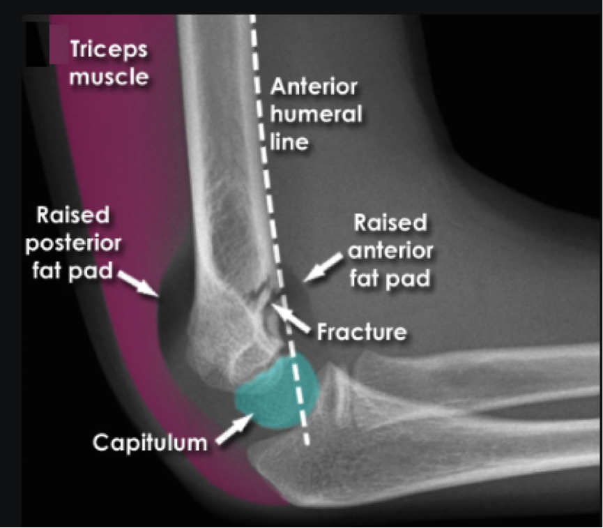
  • These abnormalities without obvious sign of fracture along bones indicative of type 1 SC fx

Dispo:

  • Type 1: long arm posterior splint, ortho follow up

  • Type 2/3: OR with ortho for reduction (closed vs. open) and pinning


 · 

Electrical Storm

storm.jpeg

Electrical Storm


Background

  • three or more episodes of sustained ventricular tachycardia, ventricular fibrillation, or appropriate ICD shocks in a 24 hour period

  • frequently, hemodynamic unstable

  • believed to mostly be due to catecholamine surge (sympathetic overdrive), but need to consider the causes below:

    • MI

    • Electrolytes

    • Acute HF

    • QT prolongation/shortening

    • Torsades

    • Brugada

    • Thyroid storm

    • Drugs

    • Sepsis

  • Presentation is broad

    • May complain of pain from ICD shocks, palpitations, syncope

    • Can present in cardiac arrest

Management

  • ABCs

  • ACLS guidelines should be followed

    • Pulse (VT)==>cardiovert

    • Pulseless (VT/VF)==>defibrillate

      1. Consider dual defibrillation if VT/VF persists after 5 delivered shocks (see image below)

        1. Coordinate firing of both defibrillators at the same time

dual defib.png

  • Should you place a magnet over ICD if patient has one?

    • Remember, magnets turn of the ability to defibrillate, but don't affect pacing capability

    • If you want the patient to be shocked then do not place a magnet, unless you think you’re dealing with something other than VF/VT and thus it is shocking inappropriately (sinus tach, afib)

      1. Practice variation exists. Some will place the magnet, especially if the pt is stable, to spare pt more anxiety/pain contributing to the storm

  • Start them on an anti-arrhythmic

    • Amiodarone 300mgè150mg IV //

    • Procainamide 10 mg/kg IV over 20 min //

    • Lidocane 1-1.5mg/kg IV

  • Add a Beta Blocker to suppress the sympathetic tone and increase the dysrhythmia threshold

    • Metoprolol 2.5-5mg IV q2-5min //

    • Propranolol .15mg/kg IV over 10 min followed by standing order//

    • Esmolol 300-500 mcg/kg push followed by drip

  • Consider an anxiolytic or sedation

  • Brugada

    • Unlike aforementioned, VF in these pts is thought to be due to excessive vagal tone

      1. Isoproterenol drip will increase the sympathetic tone

    • Quinidine has been shown to help

  • Torsades

    • Magnesium, replete electrolytes

    • If have episodes of bradycardia then add isoproterenol drip

Dispo

  • Admit to CCU if possible

  • May need cath lab, ablation, ECMO

LITFL

First10EM

 · 

Unstable Bradycardia

Unstable Bradycardia

Background

·       HR less than 60 BPM

·       Not all bradycardia is bad and scary, some presentations are benign and asymptomatic

·       Broad differential, some examples include:

  • Cardiac (structural/EP): AV block, STEMI, CHD, CM, Aortic dissection, etc.

  • Respiratory: Hypoxia, resp failure

  • Infectious: Myocarditis (viral), Lymes dx, etc.

  • Metabolic/Endocrine: electrolytes, hypothyroidism/myxedema coma, hypoglycemia, hypothermia, heat exhaustion/stroke

  • Tox/iatrogenic: BB, CCB, Dig, Clonidine, Opioids, TCAs, Keppra, Amio, etc.

  • Trauma: increased ICP, spinal injury

·       Place pacer/Zoll pads on patient

·       ACLS guidelines for bradycardia exist

Unstable patient

·       ABCs

·       Don’t let the patient D.I.E

  • Think drugs, ischemia, electrolytes

·       Place on monitor and obtain access

  • If cant get an pIV, then try IO or central line

  • *send for labs, VBG/BMP asap. Looking for electrolyte derangements (hyperK) that can change management

·       Place transcutaneous pacer pads so you’re ready to pace at any moment

  • Anterior/posterior position best

·       Try atropine

  • Doesn’t hurt to try, may work

  • .5mg IV, can be repeated q3min to a max dose of 3mg

·  If atropine is not working, high probability it wont, then start transcutaneous pacing while getting chronotropic medication (pressors) ready and then titrating to desired effect

  • Place dial on pacer mode

  • Set pacer rate >30BPM above pts intrisic rhythm (usually 60-80 BPM)

  • Set mAmp on 40, increase by 5mAmp as needed

  • Monitor for capture

    • Electric capture: downward pacer spike followed by wide QRS

    • Mechanical capture: palpate pulse and correlate with monitor/pulse ox

  • Try to give pt something for pain

  • Get ready to place a TVP

·       Pressors

  • “For symptomatic bradycardia or unstable bradycardia IV infusion a chronotropic agent (dopamine & epinephrine) is now recommended as an equally effective alternative to external pacing when atropine is ineffective.”

    • Thought: takes time to draw up meds/titrate. start external pacing. Get meds ready and administer, especially if hypotensive.

  • Can start either Epinephrine 2-20 micrograms/min or Dopamine 2-20 micrograms/kg/min, titrate accordingly

    • If epi/dopamine don’t work separately, then try them together

      • If bradycardia still not improving, then try isoproterenol 2-10mcg/min

        • Isoproterenol is an analog of epinephrine

·       Additional medications to consider

  • Digoxin  

    • Send dig level

    • Consider digibind

  • CCB

    • Calcium gluconate, high dose insulin

  • BB

    • Glucagon, high dose insuli

  • Organophosphates

    • Atropine, pralidxime

·       STEMI?

  • Usually inferior wall MI

  • Advocate for pt to go to cath lab


bradycardia.png
 ·