POTD: Foreign bodies, Ears!

potd kid ear.png

This is a two part series for POTD. Foreign bodies: Ears and Nose! Today, Ears!

potd anatomy ear.png

Quick Anatomy review to help locate that FB:

•        Anatomy

–       medial 2/3 is fixed in temporal bone –where many FBs are lodged and/or where trauma

•        Ask yourself: is it graspable or non-graspable?

–       Graspable: 64% success rate, 14% complication rate

–       Non-graspable: 45% success rate, 70% complication rate

•        What instrument/method should I use for what?

–       Alligator forceps: think something graspable like paper, foam

–       Suction tip: think something non graspable like a round object such as a bead

–       Irrigation: think something non graspable like a bead (note: do not irrigate organic material as will swell or break apart)

–       Glue: something non graspable like a bead or organic material that might swell or break if irrigated


Pearls on insect FB:

·       Kill it first. They will fight.

-        What to use? Lidocaine jelly, viscous lidocaine (2%), lidocaine solution, isopropyl alcohol, or mineral oil.

-        After they are dead, you can remove or can send to ENT for removal (most patients will want it out, can you blame them?)

o    An ENT friend of mine says to keep the insect in the ear and let them remove because we tend to cause trauma. Something to keep in mind.


What if I caused or the FB (like that insect fighting for their life) caused local trauma?

•        TM rupture?

–       Keep dry

•        When to use otic abx drops

–       Any trauma or dirty FB injury (think: that insect crawling around) or canal lacerations/abrasions.

–       What to give? Ofloxacin drops or the very expensive ciprodex.

•        ENT f/u


Pitfalls

•        Inspect after removal

–       Something else in there? Abrasions/trauma and need prophylactic antibiotic ear drops

•        If at first you don’t succeed, try again. But consider changing the technique of removal. Remember the law of diminishing returns.


References:

Pem playbook: excellent peds podcast by Dr.  T Horeczko - ‎2015

Wiki EM: Ear foreign body

 · 
Share

POTD Trauma Tuesday: Name that fracture!

A 36-year-old man presents by ambulance following a motorcycle crash. He told the EMTs that he lost control and fell sideways, bracing his fall with his outstretched right hand. His R arm looks deformed but is neurovascularly intact. An xray is obtained.

xray potd.png


What’s the name of this one again?!

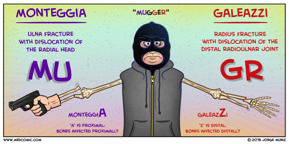
Galeazzi fracture! Named after an Italian surgeon from Milan.

What is it? a fracture-dislocation of the distal third of the radius associated with dislocation-subluxation of the distal radial ulnar joint, or DRUJ.

Why do we care? With Galeazzi fractures, there is a high risk of malunion, loss of function, infection, and chronic pain in adult patients. For this reason, surgical management with internal fixation and possible open reduction is required. The repair should occur promptly, so the emergency physician or another clinician should contact the orthopedic consultant emergently to coordinate care.

What about in peds? Emergent orthopedic consultation is still required but interestingly, in children, some Galeazzi fractures are treated conservatively with closed reduction by an orthopedic surgeon. Disruption of the DRUJ can be subtle, so a high suspicion should be maintained when a patient presents with a fracture of the distal third of the radius.

Pearls of the Peal:

* Look for fracture-dislocation of the distal radius and ulna after a fall onto an outstretched arm. This injury can’t be missed: it requires immediate orthopedic involvement.

* Skin tenting associated with the Galeazzi fracture-dislocation puts the patient at risk for skin necrosis and conversion to an open fracture.

Wasn’t there some way to remember this compared to other one?? Why, yes! See below:


mugger.png

Sources:

Comic: Medcomic.com

Xray and clinical information: PEER IX

 · 
Share

POTD: Straight leg test. A leg up on clinical testing!

A little background:

Lumbar disc herniation is the most common cause of lumbar radiculopathy, or sciatica, a shooting or burning pain from the low back radiating down the posterior leg distal to the knee.

Two tests used to evaluate these symptoms are

The straight leg raise.

·       The straight leg raise test is highly sensitive but not very specific for disc herniation.

·       This is performed by lifting the leg affected by the radiating pain.

·       The patient lies supine with one leg either straight or flexed at the knee with the sole of the foot flat on the stretcher.

·       The examiner then raises the affected leg up, extended, to 30 to 70 degrees.

·       Reproduction of low back pain that radiates down the posterior affected leg at least past the knee is considered a positive result. Not just pain to the lower back, which is a common misconception.

·       The SLR test can also be performed with the patient in a sitting position, by stretching the sciatic nerve by extending the knee; the test is positive if pain radiates to below the knee.


The crossed straight leg raise.

·       It is highly specific (90%) for disc herniation

·       You perform the same test as the straight leg but on the unaffected leg.

·       A positive test: reproducing both the back pain + the radiation down the affected leg.

Sources: Peer IX, Tintinelli’s, Dr. Sergey Motov, Uptodate

 · 
Share