POTD: Eye'm Scared!

Does anyone else get freaked out by stuff involving the eye? Well, not after this POTD you won’t.

Today I’m going to cover eyelid lacerations, probably one of the trickier ones we can encounter in the ED. First off, you must rule out corneal injury and globe rupture. Once that has been done, you can move on to considering the repair.

Repairing eyelid lacs are within the realm of the ED physician, but only under certain conditions. If any of the following findings are present, then you should involve an ophthalmologist for definitive repair.

·      Involvement of the lid margin >1mm

·      Within 6-8mm of the medial canthus (suggesting lacrimal duct/sac involvement) – can lead to poor drainage, excessive tearing and recurrent conjunctivitis or stye!

·      Through and through lacerations (involves the tarsal plate)

·      Ptosis (suggesting levator palpebrae muscle involvement)

To repair, considering using a supraorbital block or infraorbital block depending on location. Topical LET or EMLA may be considered if applied carefully to prevent leakage into eye. Then use very fine material such as 6-0 or even 7-0 sutures. These should be removed in 5-7 days and pt should follow up with an ophthalmologist ideally.

Some cool tricks tricks of the trade:

1)  To check for lacrimal duct involvement: can instill fluorescein carefully over cornea only and place a wood’s lamp over laceration. If fluorescence in wound, that means you have lacrimal duct involvement

2)  Use Tegaderm and cut a window into it using fine scissors to approximate the size/shape of wound you want to repair. Place over area of interest and can use tissue adhesive to glue together laceration; any glue run-off will get on Tegaderm instead!

3)  Use tetracaine and then place a Morgan Lens under the lids to act as an eye shield to prevent iatrogenic globe rupture while suturing.

References

https://lacerationrepair.com/techniques/anatomic-regions/lacerations-around-the-eye/

https://wikem.org/wiki/Eyelid_laceration

 · 

POTD: Trauma Tuesday - Vasopressors in hemorrhagic shock

Background pathophysiology:

Hemorrhagic shock is initially driven by a sympatho-excitatory phase attempting to compensate for acute blood loss and is characterized by vasoconstriction, tachycardia, and preserved MAP. The hypotension that subsequently follows is a result of decreased sympathetic nervous system activity from a physiologic exhaustion of endogenous catecholamines (norepi, epi) and other adjuncts (angiotensin II and vasopressin).


The traditional teaching:

The objectives of hemodynamic resuscitation in trauma is to restore adequate intravascular volume with a balanced ratio of blood products, correct pathologic coagulopathy, and maintain organ perfusion. The use of vasopressors has been traditionally discouraged in this setting as several studies have demonstrated that it leads to adverse outcomes and increased mortality risk. Permissive hypotension is advocated based on limited data that lower SBP and MAPs will result in improved mortality. Note that ATLS does not recommend the use of vasopressors currently.


Vasopressin as the pressor of choice for trauma?

The truth is that the optimal arterial blood pressure target for resuscitation of hemorrhagic shock patients is unknown. There are no studies that have come up with a concrete goal. In order to avoid increased mortality, we have shied away from using vasopressors as adjuncts in trauma resuscitation, but we know that intuitively, persistent hypotension and hypoperfusion are associated with worse coagulopathy and organ function. Thus, it would seem prudent to reconsider this all-or-nothing strategy for something more nuanced. In one of the landmark papers that demonstrated poor outcomes from early vasopressors by Sperry et al, vasopressin was the only vasopressor that was not associated with increased mortality.


In 2019, the AVERT-Shock trial demonstrated that vasopressin administration may improve blood pressure and perfusion without worsening blood loss or increasing mortality. Vasopressin has a direct vasoconstricting effect on V1 receptors but also increases the sensitivity of the vasculature to circulating catecholamines. This is why it is often used as a second-line agent in critical care settings. Theoretically, vasopressin may augment the effects of the limited endogenous catecholamines circulating when a body is in hemorrhagic shock and avoid the deleterious effects of adding exogenous ones.


However, note that this study included a much larger proportion of penetrating trauma compared to blunt trauma, potentially limiting generalizability. While it demonstrated a robust clinical difference, it was underpowered to show a statistically significant difference in mortality. Ultimately further investigations are needed, but this paper provides a great jumping off point into how we may reach for a more balanced approach to trauma resuscitation that may include both blood products AND vasopressors when the blood products alone do not seem to be restoring perfusion.

References

https://rebelem.com/avert-shock-vasopressin-for-acute-hemorrhage/

https://emcrit.org/wp-content/uploads/2022/04/Vasopressors_in_Trauma__A_Never_Event_.13.pdf

A. Sims et al., “Effect of Low-Dose Supplementation of Arginine Vasopressin on Need for Blood Product Transfusions in Patients With Trauma and Hemorrhagic Shock: A Randomized Clinical Trial,” JAMA Surg, Aug. 2019.

 · 

VOTW: Pediatric Skull Fracture

This week’s VOTW is brought to you by the UST~

A 9 month old female infant was brought into the Pediatric ED two days after a fall from a high chair. The infant vomited once after the fall but was otherwise acting normally since then. The patient was brought to the ED 48hrs after the fall for a boggy left parietal scalp hematoma. The patient had a normal physical exam apart from the hematoma.  A POCUS was performed which showed...

Clip 1 shows an oblique disruption in the cortex of the skull, indicative of a fracture. The bones have an “overlapping” appearance. A hypoechoic hematoma is present overlying the fracture.

Image 1 shows the same fracture with relevant structures labeled.

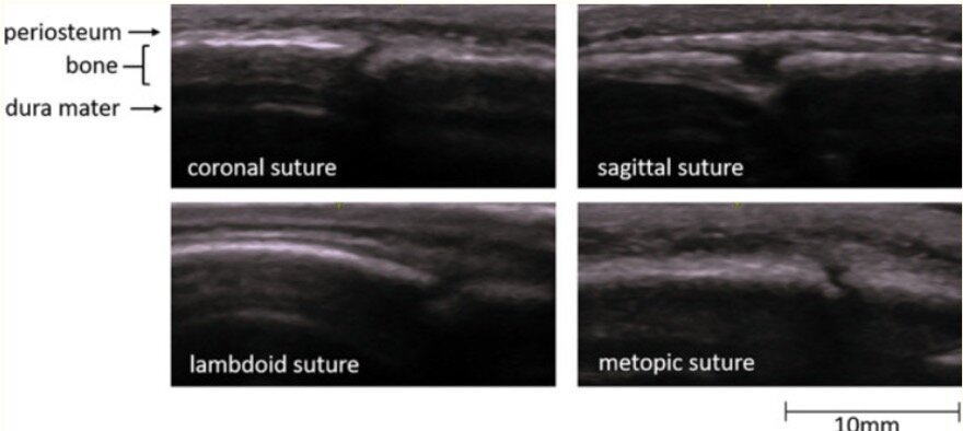
Image 2 shows a cortical disruption in the skull of the same patient, but this one is a cranial suture

Sutures and fractures look the same! How do I differentiate them?

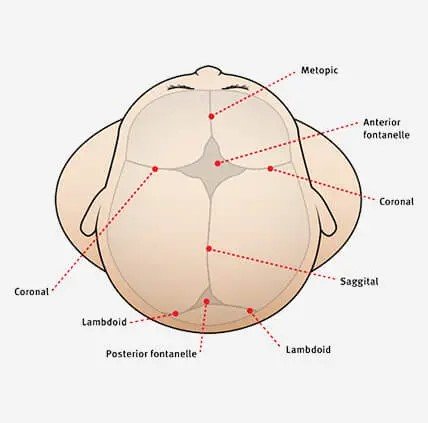
  • A suture can be followed all the way to a fontanelle.

  • Sutures are present symmetrically - scan the contralateral side if unsure

  • Fractures may appear irregular, jagged or displaced.

  • Sutures generally have an “end-to-end appearance” (image 2)- the cortex stops, there is a small space, and then restarts.

  • A fracture is likely to have an overlying hematoma.

Image 3. More examples of sutures

Image 4. A review of the anatomy of sutures and fontanelles

How to perform the study

  1. have a parent or assistant stabilize the child’s head, especially if they are squirmy

  2. use a linear high frequency probe and a lot of gel, especially if there is hair

  3. warm up the gel (put the gel bottle in your backpocket) which might make it less uncomfortable for the patient

  4. scan the area of swelling in two orthogonal planes and look for disruptions in the cortex

  5. scan the area around the hematoma as well- the fracture may not be directly under the hematoma

Clinical Decision Making

There is limited data on the use of POCUS for diagnosing pediatric skull fractures.

  •  When performed by EM Physicians, POCUS for skull fractures has sensitivities ranging from 67% - 100% and specificity of 85% - 100% (1)

  •  The presence of a skull fracture increases the likelihood of intracranial injury by four-fold (2)

POCUS for pediatric skull fractures might be most useful in the borderline case- for example a child who has an occipital/parietal/temporal scalp hematoma but otherwise looks great in the ED. Using PECARN you decide that you would rather observe this patient than subjecting the patient to radiation +/- sedation. If you decide to perform a POCUS, the absence of a skull fracture might be reassuring to you (and the family) and support your shared decision to observe the patient. The presence of a skull fracture might raise your concern for intracranial injury and change your decision about imaging. 

For a patient with a high pre-test probabiltiy for underlying pathology a negative POCUS should not be used a rule out test.

It might also be useful seeing a depressed or complex skull fracture as this may expedite imaging and specialist consultation.

More research is needed to define the role of POCUS in clinical decision making and how we might be able to integrate it with clinical decision rules like PECARN.

Happy Thanksgiving!

Your Sono Team

  1. Alexandridis G, Verschuuren EW, Rosendaal AV, Kanhai DA. Evidence base for point-of-care ultrasound (POCUS) for diagnosis of skull fractures in children: a systematic review and meta-analysis. Emerg Med J. 2022 Jan;39(1):30-36. doi: 10.1136/emermed-2020-209887. Epub 2020 Dec 3. PMID: 33273039; PMCID: PMC8717482.

  2. Kuppermann N, Holmes JF, Dayan PS, et al.. Identification of children at very low risk of clinically-important brain injuries after head trauma: a prospective cohort study