Wayne Pneumothorax Tray

I wanted to do a little blurb about the pigtail kit at Community. I often find that we as providers become pretty comfortable with what we know and uncomfortable with any tools we haven't used before. Back in July, I had to do a chest tube at Community, and the kit was totally different (and rest of the procedure was completely different because of this). This kit is not saldinger technique, and doesn't require use of needles (though you still should use lido obvs). I was initially confused when I was looking at the kit, and so wanted to write this out in case you face the same!


The kit comes with a 14Fr pigtail, trocar, long blade that goes in trocar (looks like a hollow bore needle, but isn't!), 11 blade, tubing, three way stopcock, and one way air valve. The main difference from the pigtail kits that we're used to, is there is no guidewire and no needle! Meaning, you're not going in with the needle first. 


Essentially, you will end up inserting the pigtail with trocar and long blade in one piece, into the incision site. The trocar is placed in a larger fenestrated hole towards the end of the pigtail.


The steps for the procedure include;


  1. Confirm the location, fool (pick the side with the pneumo, and do it in the triangle of safety)

  2. Prep the site with chlorhexadine

  3. Anesthetize the site with lido

  4. Get sterile

  5. Drape and re-prep (you could probably prep once, but I'm a little OCD)

  6. Combine the pigtail, trocar, and long blade as shown in image

  7. Make your incision above the rib with the 11 blade

  8. Taking the combined long blade, in trocar, in pigtail - insert at your incision, aimed towards the lung apex

  9. Remove the long blade once you pass the resistance of the pleura

  10. Advance the trocar and pigtail, before removing the trocar and continuing to advance the pigtail to the desired depth (usually around 15-20 cm)

  11. Suture the pigtail in place and place a dressing over it

  12. Attach the tubing with the one way valve or to a pleurovac


Now for those of you that may read this and say "omg, I'm not trying to just stab someone," well, you are not alone. Others have commented the same. And if you are so inclined to place this pigtail using saldinger technique, that is still possible. You will need to crack open a central line kit and pillage the needle, syringe, and guidewire. The trocar in the Wayne Pneumothroax tray is hollow bore, and the guidewire can still be fed through that. Hope this was helpful! 


Unstable Pelvic Ring Fractures

The pelvic ring consists of the sacrum and two innominate bones, which are made up of the pubis, ilium, and ischium. These bones are held together by strong ligaments to give the pelvis stability.

A pelvic ring fracture is a severe fracture with 2 breaks in the circular ring, leading to an unstable pelvis and a potentially unstable patient. Fractures that disrupt the pelvic ring predispose patients to bleeding given the large network of arterial and venous anastomoses. Patients who have an isolated pelvic fracture and are hypotensive carry a mortality of 15-40%. Most vascular injuries in the pelvis are venous (90%). While rare, arterial bleeds (10%) should be suspected when a pelvic binder is placed but the patient remains hemodynamically unstable. The retroperitoneal space can accumulate 4 liters of blood before venous tamponade occurs. Pelvic binders are useful in that they can help tamponade bleeding veins, decrease total pelvic volume, and prevent the shifting of bony fragments.

Other unstable pelvic fractures include lateral compression fractures, "open book" pelvic fractures, and vertical shear fractures. Lateral compression fractures occur when a lateral force vector (t-bone in an MVC) causes an anterior ring disruption and sacral fracture.

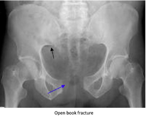
“Open book” fractures occur as a result of anteroposterior compression injury to the pelvis, commonly caused by high-speed trauma or elderly falls. There is a disruption to the pubic symphysis and the pelvis opens like a book. Diastasis of > 1 cm (blue arrow) can indicate instability. Disruption of the pubic symphysis, one of the strongest ligamentous structures in the human body, requires a lot of force and should be a red flag to look for other injuries to the head, spine, chest, or abdomen.

Vertical shear pelvic fractures are seen when one-half of the pelvis shifts upward as a result of a fracture of ipsilateral anterior and posterior pelvic ring fractures. They typically occur as a result of high-energy force applied in the axial direction (aka from the gas pedal to the femur and up to the pelvis). Patients may have an unstable pelvis and leg length discrepancy.

For all unstable fractures, you should appropriately resuscitate and stabilize the patient. Give blood as needed but avoid transfusing through lower limb access because it may drain into the retroperitoneal space. If there is a pelvic ring fracture, consider binding the pelvis. Your binder should lay over the greater trochanters and have enough force to close the pelvic ring (video:https://www.youtube.com/watch?v=tWLBZKeWEkg).


Galea Lacerations

Anatomy:

 The galea is a dense white layer that covers the periosteum of the skull. It serves as an insertion point for the frontalis and occipitalis muscles


Five layers of the scalp

·      SCALP

o   Skin

o   Dense Connective tissue

o   Aponeurosis (galea)

o   Loose connective tissue

o   Periosteum


Dense connective tissue layer is richly vascularized. The tight adhesion of these vessels to the connective tissue inhibits effective vasoconstriction, hence the large amount of bleeding in scalp lacerations.


The loose connective tissue layer = the DANGER ZONE when lacerated. This layer contains the emissary veins, which connect with the intracranial venous sinuses. Lacerations at this layer are high risk for spreading infection to the meninges!


Approach:

·      Examine the wound, clear of debris, and assess the depth of the wound.

o   Superficial wounds generally don’t gape

o   Deep wounds gape widely due to laceration of aponeurosis, and the tension from the frontalis muscle and occipitalis muscle pull the wound open in opposite directions

·      Hair removal unnecessary unless it interferes with actual closure or knot tying. No increased risk of infection if you do not remove the hair. Shaving head increases risk for infection!

·      Obtain hemostasis with pressure and lidocaine with epinephrine.

·      If the galea is lacerated more than 0.5 cm it should be repaired with 3-0 or 4-0 absorbable sutures. to prevent a serious cosmetic deformity from developing.

·      Skin can be repaired using staples; interrupted, mattress, or running sutures, such as 3-0 or 4-0 nylon sutures; or the hair apposition technique. Removal of sutures or staples in 14 days.

·      Antibiotics - With open skull fractures (blunt or penetrating), should give antibiotics: Ceftriaxone 2 grams q12hr + vancomycin for 24 hours.


Complications:

·      Asymmetric contraction of the frontalis muscle

·      Osteomyelitis, brain abscess - Failure to repair can also allow bacteria to get to the loose connective tissue layer more easily between the galea and periosteum, leading to increased risk of infection

·      Subgaleal hematoma


References:

https://sjrhem.ca/rcp-scalp-lacerations-you-can-leave-your-hat-on/

http://pemsource.org/2019/01/01/question-trauma-10/

https://aneskey.com/special-anatomic-sites/

https://www.aafp.org/afp/2017/0515/p628.html

https://www.vumc.org/trauma-and-scc/sites/default/files/public_files/Protocols/Antibiotics%20in%20CranioFacial%20Trauma%202021.pdf

Tintinallis Emergency Medicine a Comprehensive Study Guide 8th Edition

 ·