Pulse Cooximeter: how to use it

On my previous shift, we had 2 patients with a “gas” exposure at their apartment. 

So the clinical question arose: How can we rapidly screen patients with “gas” exposure? 

First, any abnormal vitals raise a red flag and automatically take us off the rapid discharge pathway. These patients need appropriate triage to (

likely

) north side. 

In any patient who has been exposed to a fire or there is concern for carbon monoxide exposure, we have the Masimo pulse-cooximeter. 

It is located in the south side charge nurse station. 

To utilize this device: hold down the power button while the pulse sensor is on the patients finger.

masimo_co-oximetry.jpg

After pressing the display button, it should look as below:

Please note that there is a continuous SpCO and SpMet in green on the left and right of the display, respectively. 

If results in an asymptomatic patient with a low risk exposure are normal, they can be safely discharged without further testing. However, in a symptomatic patient with a normal pulse-cooximetry, they should be further screened with blood gas cooximetry. Furthermore, a

ny abnormal value of %SpCO>5% should be repeated with a blood-cooximetry.

Smokers may have a baseline CO-Hgb of 5-6%, and may require confirmatory testing with blood-cooximetry through our blood gas lab. 

In short, if patient has a %SpCO <5% and is asymptomatic they may be safely discharged. This also requires a normal %SpO2 because %SpO2<85% decreases the accuracy of the Masimo pulse co-ox, as per the literature posted on their own product page. 

Final summary of this POD:

we have a pulse-cooximeter.

Utilize it for rapid screening and for reassurance of low risk patients. 

Please clean the finger sensor between patients with a purple wipe

As always: feedback both negative and positive IS STRONGLY ENCOURAGED. 

TR,

Wells 

 · 

Suction cannisters in series

Today during my TR shift, CY asked me for demonstration of how to connect suction canisters in series for large volume paracentesis.

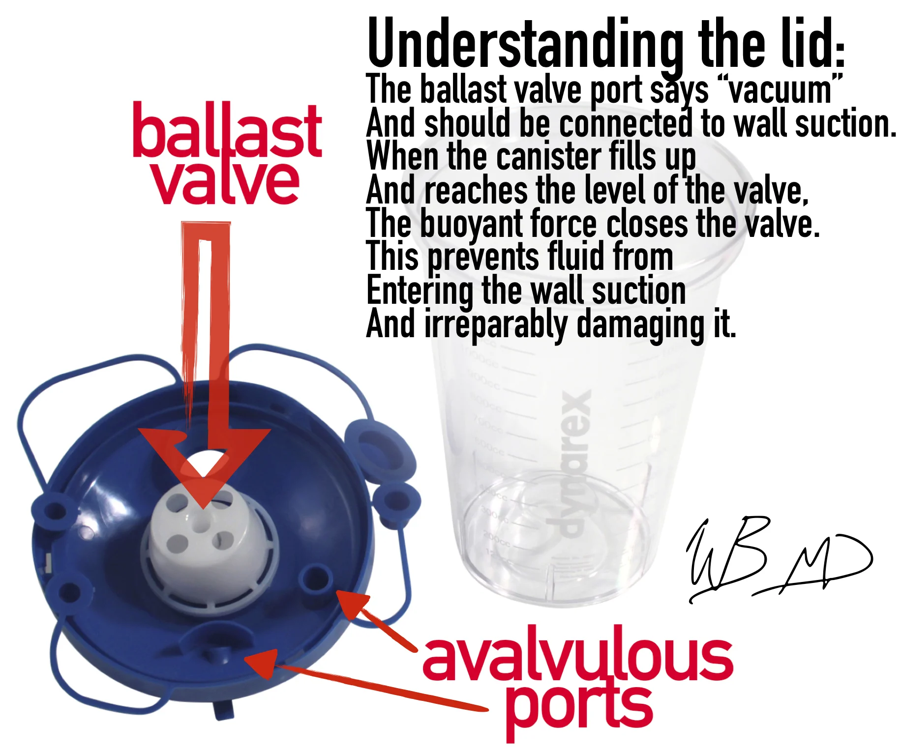
Before we go into the actual technique, lets take a second to understand how the suction canister functions. This is not our exact model of suction canister lid, but is functionally the same. See my infographic below:

suction-kanister-explained.jpg

The goal: to connect 3 of the vacuum canisters together so that when one fills, the fluid then starts draining into the next. Once the second fills it will drain into third and automatically stop.

With this technique you can perform your paracentesis, start drainage, and then safely walk away. Please instruct your patient not to move whilst drainage is in action.

So connect one suction canister with vacuum tubes as you normally would: vacuum to wall suction; the next vacuum tube should go from either of the avalvulous ports to the next avalvulous port. Critical point: the only port that needs to be connected to the ballast valve port is the port that plugs into wall suction. All subsequent ports should be connected in series with the avalvulous ports.

The final attachment is the avalvulous port to christmas tree adapter, which is included in our thora/paracentesis kit.

During what I believe was my intern year, I had shown Strayer this technique, and much to my chagrin, he tweeted out a post about it. See below:

strayers_ccannister-post-1.jpg
suction-kanister-explained.jpg

The benefits of utilizing this technique for large volume paracentesis are twofold: (1) increased safety and (2) utilization of resources (in this case physician time). Safety is increased because this method does not utilize sharp needles to transfer fluid to a vacuum flask; thereby, it reduces the risk of needle stick injury (especially important for the population of patients that need ED paracenteses). Utilization of resources is improved because a physician does not need to remain at bedside or repeatedly check the progress of evacuation, assessing for need to switch to the next vacuum flask.

For large volume paracentesis, I recommend only connecting a max of 4 of our suction canisters in series. I typically do 3, but 4 approaches that max of 5L that current thought suggests may cause hemodynamically significant fluid shifts.

 · 

Infiltration of IV contrast into SQ tissues

I think we all dread this: Just imagine this scenario: you are concerned that your patient has a pulmonary embolus. They are tachycardic, hypoxic on room air, but BP’s are stable (for now). They were a tough stick and 2 nurses tried for 5 minutes, so under real time ultrasound guidance you place what you think is an expert 20g x 48mm angiocath in their right cephalic vein.

You dial 6308 and ask the radiology technologist to scan the patient next, then you are off to see the next patient on a busy north side shift.

15 minutes later, you get a call: “Ummmm… Doc, patient x…. that line blew. They got the hole load in the right arm. Can you come take a look?”

This can happen about 0.24% of patients who receive IV contrast in the upper extremity (or about 1/400 studies).

UNDERSTANDING CONTRAST:

The Omnipaque™ we use for our IV contrast CT studies is Iohexol:

Here is its structure:

1200px-Iohexol.svg_-1.png

The concentration we use is 350 mg/mL, this results in a total osmolality of 844 mOsm/kg water. This is approximately 3 times the osmolality of serum. However, given the fact it is non-ionic, it does not cause a significant osmolar effect. Nonetheless, infiltration will lead to fluid shift of H2O down its gradient into the tissue compartment the infiltrated contrast material occupies, which is typically the interstitial space. This has been shown experimentally in a renal model: after contrast load urine output increases because the iohexol is renally excreted.

The most danger for infiltration occurs with high flow rates achieved during CT angiography studies. As per our technologist on our Siemens machine, the auto injector we use has a flow rate 4-5 mL/s achieved with a pressure gradient anywhere from 50-100 psi. If the IV has poor flow due high intracatheter or intravenous resistance, then the pressure can increase to upward of 400 PSI to achieve flow. When this high pressure occurs, the IV can become dislodged. Remember Newtonian mechanics: Pressure exerts a force, and for each force there is an equal and opposite reaction.

MANAGEMENT OF IV INFILTRATION OF A CONTRAST LOAD:

####first and foremost, you need to inform the patient what happened. Explain why this happened. Apologize for the complication. Address any pain issues. Tell them how you and the team will care for their likely painful upper extremity. Finally, you can reassure them for the reasons below.

*Gradually, the infiltrated contrast will diffuse out of the tissue compartment and back into the serum. This diffusion can be enhanced with gentle warm compresses.

**Plastic surgery recommendations for this complication are elevation of the extremity, local massage, and frequent neuromuscular checks.

***In a study of 102 cases of IV contrast extravasation in the upper extermity, surgical intervention was required in 0. The surgeons behind this study recommend surgical consultation if there obvious signs of skin compromise or compartment syndrome.

****compartment syndrome has been reported in the case literature, so please, please, please, watch for that. The incidence is on case report level, so it is likely very, very low.

SOME TIPS PREVENTION OF IV INFILTRATION OF US GUIDED PIV’s:

*Always use the 48 mm angiocath

**The more of the catheter that is in the lumen of the vein, the less likely it is to become dislodged. So a good rule of thumb is at least 1 cm of the catheter within the lumen of the vein. So any target >2cm deep to the skin will be difficult to properly position using a 45 degree angle of approach.

One fun final fact: non-ionic iodinate contrast materials are procoagulants and as per the FDA they should be avoided at all cost patients with homocystinuria.

AS ALWAYS FEEDBACK IS ENCOURAGED.IF YOU HAVE AN INTERESTING QUESTION, FACTOID, OR OPINION, PLEASE RESPOND ALL.

Best regards,

Wells

References:

https://www.accessdata.fda.gov/drugsatfda_docs/label/2015/018956s095,020608s031lbl.pdf

https://www.sigmaaldrich.com/catalog/substance/iohexol821146610895011?lang=en®ion=US

Jonas Pologe’s US PIV POD

Belzunegui T, Louis CJ, Torrededia L, Oteiza J. Extravasation of radiographic contrast material and compartment syndrome in the hand: a case report. Scand J Trauma Resusc Emerg Med. 2011;19:9.

Sbitany H, Koltz PF, Mays C, Girotto JA, Langstein HN. CT contrast extravasation in the upper extremity: strategies for management. Int J Surg. 2010;8(5):384-6.

Solomon R. Contrast media: are there differences in nephrotoxicity among contrast media?. Biomed Res Int. 2014;2014:934947.

 ·